Maxillofacial trauma is typically caused by blunt trauma due to interpersonal violence, moving vehicle accidents (MVA), falls, or sporting activities 1. Because these activities transcend cultural and geographic borders, maxillofacial trauma remains a global health issue. Technological advances have resulted in faster methods of transportation and with these developments maxillofacial trauma has become a major public health issue also in the developing world. Although management of the airway and cardiopulmonary system take precedence, facial injuries demand attention because of their potential for long-term impact on quality of life (QOL) and the patient’s psychological wellbeing2 as the face houses many vital structures necessary for sight, hearing, speech, olfaction, and mastication.
When energy transferred to a bone exceeds its stress tolerance, a fracture may result. Several factors determine if and how a bone fractures: the amount of energy transferred to the bone e.g. mechanism of injury, speed of collision, airbag deployment etc.; vectors of forces; and the characteristics of the tissue involved e.g. anato-ic location of impact and quality/health of the bone.
A simple fracture occurs when a single site of discontinuity exists between two bony segments. With a comminuted fracture there are multiple segments of bone. Bone segments protruding through skin are considered open fractures, as are fractures of the alveolar bone and sinuses. They are more susceptible to infection compared to their closed counterparts. Closed fractures are uncommon in the face.
The bony fragments may remain anatomically reduced or can become displaced depending on the mechanism of injury and muscular forces acting on the fragments. The degree of anatomic disruption of fractured segments is referred to as displacement. Blood flow to the fracture site results in callus formation over time followed by mineralisation of the callus if the fracture site is immobilised.
Malunion occurs when fracture segments are not reduced and the bone heals in an anatomically incorrect position3. In the face, this can cause significant disfigurement and disability.
Disfigurement occurs because the skin soft tissue envelope (SSTE) of the face is dependent upon the underlying maxillofacial skeletal framework. Anatomic perturbations of the bony framework from maxillofacial trauma manifest as abnormal appearances of the SSTE. Depending on the location of the fracture(s), the aesthetic deformity may be inconspicuous e.g. minimally displaced anterior maxillary sinus wall fractures, or obvious e.g. displaced nasal bone fractures.
Disability occurs when malunion interferes with function. This is most evident with dentoalveolar or mandibular fractures, as malunion can result in dental malocclusion, making mastication difficult or impossible.
The surgeon has to ensure that bony fragments are anatomically reduced and stabilised so that when healing is complete, callus formation is minimised to minimise disfigurement and disability. Even when reduction is achieved, the result may not be ideal. Poor blood supply or wound infection may cause delayed union when bone fails to mineralise 3 months after reduction and immobilisation. Fibrous union may occur (usually within 10 days in adults) if bones are reduced but inadequately immobilised. Nonunion may result if bone is lost or the wound is contaminated with foreign material. Therefore, the surgeon must ensure that a fracture site is clean, free of foreign material (insomuch as possible), and well vascularised prior to reduction and immobilisation. Antibiotics are sometimes warranted to counter bacterial contamination.
Not all maxillofacial fractures require intervention. The primary goals of repair of craniomaxillofacial (CMF) fractures are restoration of premorbid anatomy and function:
Fracture reduction and fixation are best performed in the 1st two weeks following injury 5. Strict attention to the following principles optimise bone healing and surgical outcomes
Anatomic reduction of fractured segments requires a 3-dimensional understanding of maxillofacial anatomy. Nonetheless, substantial individual variation is common, and premorbid photographs or examination of the unaffected side of the face can be beneficial. Intraoperative imaging is also used by some to achieve adequate reduction5, although such technology is costly and time consuming. Bilateral fractures may preclude using the contralateral face as a template. In many cases, the surgeon must reference normal anatomic landmarks. However, when there are no anatomically normal bony landmarks to use as reference, one should first restore dental occlusion, followed by the buttresses of the face (see section on maxillo-mandibular fixation and anatomy).
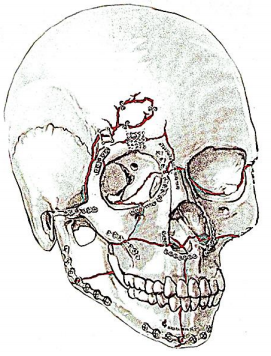
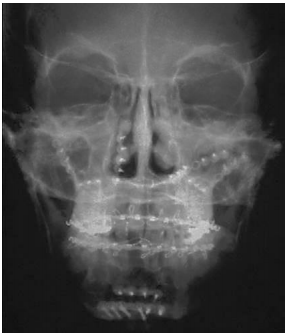
Fixation of bone across fracture sites is done to immobilise the bone against musculoskeletal forces and to permit mineralisation to occur. Fixation is generally accomplished using titanium plates secured with screws (Figures 1, 2, 3, 4).
energy transferred to a bone exceeds its stress tolerance, a fracture may result. Several factors determine if and how a bone fractures: the amount of energy transferred to the bone e.g. mechanism of injury, speed of collision, airbag deployment etc.; vectors of forces; and the characteristics of the tissue involved e.g. anatoic location of impact and quality/health of the bone.
A simple fracture occurs when a single site of discontinuity exists between two bony segments. With a comminuted fracture there are multiple segments of bone. Bone segments protruding through skin are considered open fractures, as are fractures of the alveolar bone and sinuses. They are more susceptible to infection compared to their closed counterparts. Closed fractures are uncommon in the face.
The bony fragments may remain anatomically reduced or can become displaced depending on the mechanism of injury and muscular forces acting on the fragments. The degree of anatomic disruption of fractured segments is referred to as displacement. Blood flow to the fracture site results in callus formation over time followed by mineralisation of the callus if the fracture site is immobilised.
Malunion occurs when fracture segments are not reduced and the bone heals in an anatomically incorrect position3. In the face, this can cause significant disfigurement and disability.
Disfigurement occurs because the skin soft tissue envelope (SSTE) of the face is dependent upon the underlying maxillofacial skeletal framework. Anatomic perturbations of the bony framework from maxillofacial trauma manifest as abnormal appearances of the SSTE. Depending on the location of the fracture(s), the aesthetic deformity may be inconspicuous e.g. minimally displaced anterior maxillary sinus wall fractures, or obvious e.g. displaced nasal bone fractures.
Disability occurs when malunion interferes with function. This is most evident with dentoalveolar or mandibular fractures, as malunion can result in dental malocclusion, making mastication difficult or impossible.
The surgeon has to ensure that bony fragments are anatomically reduced and stabilised so that when healing is complete, callus formation is minimised to minimise disfigurement and disability. Even when reduction is achieved, the result may not be ideal. Poor blood supply or wound infection may cause delayed union when bone fails to mineralise 3 months after reduction and immobilisation. Fibrous union may occur (usually within 10 days in adults) if bones are reduced but inadequately immobilised. Nonunion may result if bone is lost or the wound is contaminated with foreign material. Therefore, the surgeon must ensure that a fracture site is clean, free of foreign material (insomuch as possible), and well vascularised prior to reduction and immobilisation. Antibiotics are sometimes warranted to counter bacterial contamination.
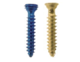
Screws are either self-tapping or self-drilling. Self-tapping screws require pre-drilling prior to inserting the screws, but also require less force which is advantageous when fixing fragile bony segments. Larger plates and screws provide greater stability and immobilisation. Non-locking plates are typically used for the midface. These plates must be shaped to fit the bone precisely or they will distract the bony segment towards the plate when the screw is engaged.
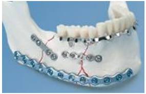
Locking plates can be helpful when immobilising mandibular fractures. When used with locking head screws, locking plates do not require exact coaptation of the plate to the bone as the bone is not compressed against the plate (Figures 3, 4). This avoids impairing cortical bone perfusion, and provides a more stable fixation when compared to a non-locking system.
Because titanium plates and screws are too expensive for many developing world centres, reliance is placed on stainless steel wire to reduce and fix fractures.
Preventing infection begins with initial management in the emergency unit. Open wounds are copiously irrigated with sterile isotonic fluid. Badly contaminated wounds are thoroughly cleaned and irrigated. Pressurised pulsatile irrigation methods have been demonstrated to be useful in wound debridement 6 and are particularly useful to cleanse wounds with particulate matter e.g. sand and road debris. Prophylactic antibiotics to prevent infection when treating non-mandibular maxillofacial fractures have been discouraged 7, although a recent review highlighted the lack of large high-quality randomised controlled trials to evaluate antibiotic use in non-mandibular facial fractures. When treating fractures of the mandible, prophylactic antibiotics are likely beneficial when used in the immediate perioperative period 8. In any case, adherence to sterile technique in the operating room and good quality postoperative wound care should not be compromised.
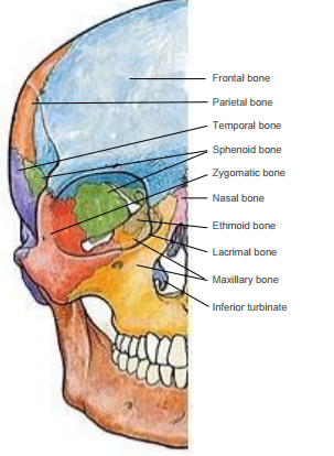
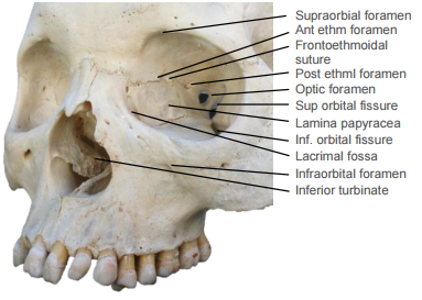
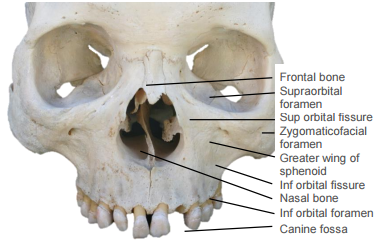
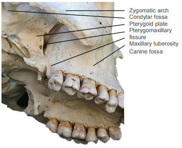
Knowing which fractures require treatment requires a thorough understanding of the anatomy of the craniomaxillofacial (CMF) skeleton and how forces are transmitted (Figures 5-9). This anatomical knowledge, coupled with a detailed history, physical examination, and imaging permit the surgeon to establish a treatment plan for various fracture patterns.
Because of the complexity of the CMF skeleton, it is helpful to divide injuries into the following anatomic regions: frontal sinus, nasoethmoid complex (NEC), zygomaticomaxillary complex (ZMC), midface, palatoalveolar, and mandible.
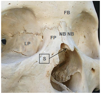
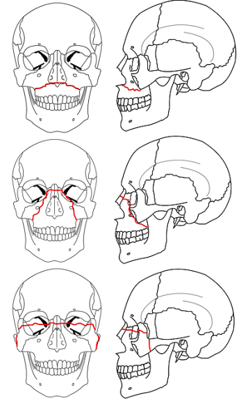
Four paired, vertically-oriented bony pillars (buttresses) i.e. the nasomaxillary (NM), zygomaticomaxillary (ZM), and pterygomaxillary (PM) buttresses 10, and the vertical rami of the mandible support the sinus cavities and give structure to the craniofacial skeleton (Figure 10). They resist deformation by the powerful forces of mastication and other vertical forces 9.
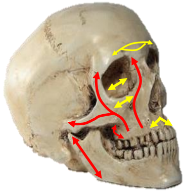
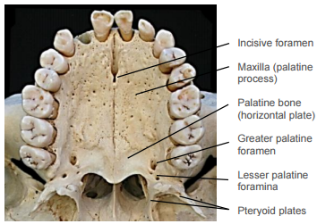
The NM buttress extends from the nasion inferiorly along the pyriform aperture. The ZM buttress extends from the frontozygomatic suture line inferiorly through zygomaticomaxillary suture line to the maxillary alveolus. The PM buttress extends from the cranial base superiorly to the inferior border of the pterygoid plates. Transversely oriented buttresses include the frontal bar, infraorbital rim and nasal bones, and the hard palate and maxillary alveolus (Figure 10).
It is helpful to think of the face as skin and soft tissue draped over the sinus cavities which are enclosed by thin bone; hence the appearance and structure of the skin depends on the structure of the underlying craniofacial skeleton and the integrity of the buttresses. Repair of maxillofacial injuries is therefore directed at reestablishing relationships between the buttresses and surrounding bone and soft tissue, as well as reduction and stabilisation of the integrity of the buttress(es).
History
The craniofacial trauma surgeon generally remains uninvolved with patient care in the emergency department until stabilisation of the airway and cardiopulmonary systems is complete. Nonetheless, it is certainly possible that craniofacial trauma may compromise the airway or vascular system in which case the experience of a head and neck surgeon can be particularly helpful. In such scenarios, an abbreviated history can be obtained while surgical intervention is taking place in the emergency department. For instance, a patient with bilateral mandibular fractures may present in air-way distress because of posterior displacement of the oropharyngeal tongue. In such a case, anterior traction on the tongue using a penetrating towel clamp or by digitally grasping the tongue with a gauze swab can facilitate endotracheal intubation. In some cases, endotracheal intubation may be difficult or precluded (because of suspected laryngeal injury), necessitating tracheostomy or cricothyroidotomy. In cases of vascular compromise, exanguination is a risk. Typically, tamponade is crucial for haemostasis in emergency situations until formal wound exploration and repair can be completed. Once the airway, breathing, and circulatory systems have been stabilised, a more thorough history can be obtained.
The surgeon should determine the mechanism of injury i.e. fall, assault, moving vehicle collision, and the speed of the injury. While treatment of the patient can continue without knowing the exact mechanism of injury, information regarding mechanism can help the clinician understand the injury pattern and possibly reveal more inconspicuous injuries. For instance, a patient involved in a high-speed motor vehicle collision will typically be at higher risk for more severe injuries to the craniofacial skeleton. Knowing this information may encourage a clinician to order more comprenhensive imaging studies.
In addition to the mechanism of injury, the time and location of the injury can be important. These factors can provide clues regarding the status of the patient’s wounds i.e. presence of debris, risk of infection, etc. It is also crucial to ascertain the condition of the patient when the first respondents arrived e.g. was the patient unconscious, cooperative, or combative? This information can provide clues regarding the neurologic status of the patient. The Glascow Coma Scale (GCS) upon arrival of the first responders is information typically presented to the trauma surgeon.
It is not uncommon that patients with craniofacial injuries were under the influence of alcohol or drugs when they sustained an injury. This information should also be obtained.
In an awake and appropriately responsive patient, it is helpful to obtain a complete history directly from the patient as well.
Important questions to ask include:
Once the patient’s complaints have been documented, a complete past medical and surgical history should be obtained. The surgeon should be aware of the status of the cervical spine, which may preclude positioning changes in the operating room. Additionally, a list of current medications, allergies, family history and review of systems should be obtained. The patient’s tobacco, alcohol, and illicit drug use history should be noted.
A complete head and neck examination is performed. This includes a neurological examination which begins with assessment of the patient’s sense of person, place, and time. Cranial nerves II-XII are examined. Visual actuity, pupillary light reflexes, and extraocular movements are assessed. Facial sensation should be noted in all three divisions of the Vn. The motor division of the Vn is examined by asking the patient to bite down while palpating the masseter muscle. Facial nerve function should be carefully examined by asking the patient to raise the eyebrows, close the eyes tightly, smile, purse lips, puff cheeks, and depress the lower lip. The cochlear division of the VIIn can be grossly assessed using a conversational voice, and more detailed information regarding audiologic status can be obtained with a tuning fork exam. The IXn and Xn can be tested with the gag reflex and the voice quality. XIn function can be examined via shoulder shrug and head turn. The XIIn can be examined by assessment of tongue movement.
The scalp should be carefully examined for lacerations, foreign debris, or haematomas.
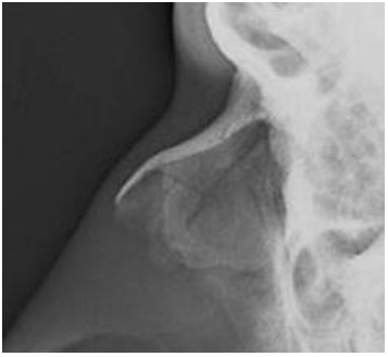
Otoscopy is essential to rule out external auditory canal lacerations, which can result in canal stenosis if left untreated. The tympanic membrane should be visualised to rule out haemotympanum, which may be an indicator of a temporal bone fracture. Clear or thin bloody otorrhoea may be found due to cerebrospinal fluid (CSF) leakage. In such cases, fluid should be collected and sent for beta-2 transferrin analysis. The mastoid bone should then be examined for Battle’s sign (Figure 11) along with the auricle.

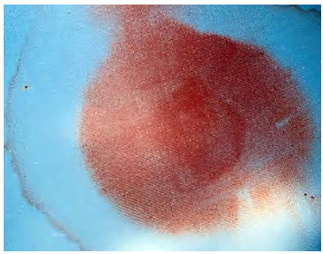
Nasal examination is important to rule out obvious fractures. Anterior rhinoscopy should then be performed to rule out septal hematoma or epistaxis. Septal haematoma requires immediate drainage to prevent abscess formation and/or cartilage destruction, which can result in severe nasal disfigurement. Open fractures of the nasal septum can sometimes be found on anterior rhinoscopy. The surgeon should also assess for watery rhinorrhea, which may be indicitive of CSF leakage. Beta-2 transferrin testing can confirm the identity of the fluid. When unavailable, the halo test can sometimes be helpful. A drop of the fluid should be placed onto a gauze swab. Blood coalesces into the center, leaving a ring around the blood in the presence of CSF (Figure 12). If deemed necessary, nasendoscopy can also be performed to further examine for CSF leakage.
Examination of the oral cavity begins with assessment of occlusion. While all patients will not have Angle Class I occlusion, it is imperative to assess if the patient’s occlusion has changed since the accident. A gross dental exam is also helpful to determine if oral-maxillofacial surgery consulation is required. The maxilla should be grasped and assessed to determine stability. The oral cavity is then examined with attention to the buccal mucosa, Stensen’s duct, floor of mouth, tongue, palate, and maxillary/mandibular alveolar ridges. The oropharynx is then examined. If indicated, nasolaryngoscopy can be performed to examine the larynx. This is required if the patient presents with otherwise unexplained respiratory distress, voice changes, or crepitus.
The facial soft tissues are then carefully examined to rule out lacerations and abrasions. The underlying bony skeleton can be palpated to assess for step-offs.
The neck should then be examined. Palpation can reveal haematoma, crepitus, or displacement of the airway. Cervical spine status is essential to know prior to manipulating the patient’s head and/or neck.
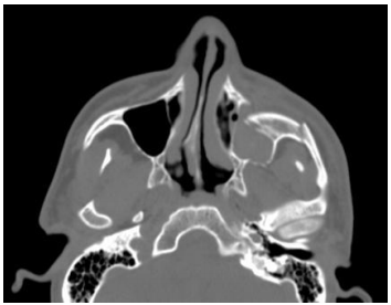
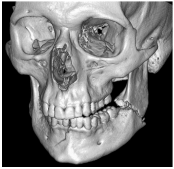
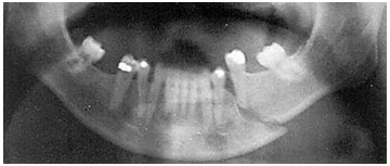
Imaging may be required to exclude injury to the brain, vasculature, and cervical spine and other associated injuries. Standard modern-day imaging for CMF skeleton includes fine-cut, uncontrasted computed tomography (CT) of the CMF skeleton (Figure 13). It is often helpful to obtain coronal reformats and three-dimensional reconstructions (Figure 14). In the absence of CT imaging, reliance is placed on plain X-rays; panorex images are helpful with mandibular fractures (Figure 15). When available, the ideal imaging study of choice for maxillofacial trauma is non-contrast CT scan of the face with fine cuts, coronal and sagittal reformats, and 3D reconstructions.
The most important consideration when reducing facial fractures involving or alter-ing the occlusal surface is to ensure that premorbid dental occlusion is restored.
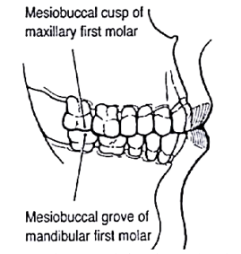
Angle class I occlusion is considered normal. It is defined as intercuspation of the mesial buccal cusp of the 1st maxillary molar with the buccal groove of the 1st mandibular molar (Figure 16). The maxillary incisors should extend slightly anteriorly past the mandibular incisors, known as overjet. The amount of overlap between the labial surface of the mandibular incisor and the lingual surface of the maxillary incisor is termed overbite (Figure 17).
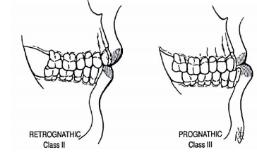
Angle class II (retrognathic) and Angle class III (prognathic) occlusions describe alternative relationships. Angle’s occlusal classification does not address anterior or transverse dental relationships (Figure 18).
One to 3mm of overjet and overbite is considered normal; if excessive, it is considered to be a form of malocclusion. Transversely, the buccal cusps of the mandibular molars should lie between the buccal and lingual cusps of the maxillary molars (Figure 16).
Stepwise treatment of fractures
Treatment of facial fractures should not necessarily be tailored to achieve Class I occlusion; rather, the goal should be to restore premorbid occlusion. Premorbid occlusion is determined by assessing the positions of wear facets on the dentition intraoperatively.
Prior to fixation of fracture sites, appropriate reduction is achieved and the patient is placed into mandibulomaxillary fixation (MMF) using arch bars, bone supported devices or with direct interdental wiring techniques 11 (Figure 19). For a detailed description of arch bar placement, readers are referred to the cited URL 4.
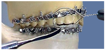
Arch bars can often be removed if adequate fixation has been achieved with plating. However, if arch bars have been used as a tension band, they should remain in place for 6 weeks.
Exposing fractures
Once the patient has been placed into MMF (if required), the fracture sites are exposed. Several approaches are available to access the facial skeleton depending on the site and extent of the fractures. Lacerations overlying the fractures are utilised to access the underlying bone if possible.
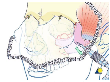
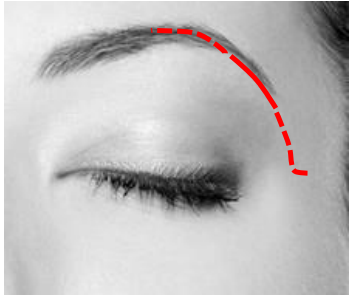
These can be addressed using a coronal incision (Figure 20). The incision extends from just superior to the root of the helix, and is carried across the parietal bone keeping behind the hairline of the scalp, and ending just superior to the contralateral helical root. Medially the incision may be extended to the pericranium if a pericranial flap is planned, or onto the cranium; laterally the dissection is typically kept in a plane just deep to the temporoparietal fascia with special care taken when approaching the zygoma to avoid the frontal branch of the facial nerve which resides in this plane (Figure 21)
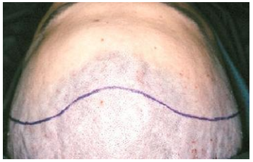
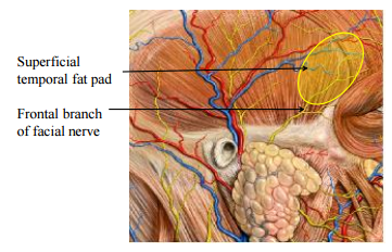
Figure 22 illustrates the coronal flap reflected anteriorly, taking care to preserve the supraorbital nerves if possible.
Access to the lateral orbital rim and frontozygomatic suture can be achieved by lateral brow or lateral lid crease incisions (Figure 23). Oftentimes isolated fractures of this region can be fixed using a mini-plate with at least 2 screws on each side of the fracture line.
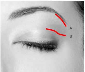
The lateral brow approach provides direct access to the superolateral orbital rim. No neurovascular structures are at risk. Scarring is generally concealed if the incision is placed within the hairs of the brow although it can cause alopecia if not carefully placed. It is used for fractures high up on the lateral orbital rim.
The lateral lid crease approach provides direct access to the lateral orbital rim (Figure 23).
Fractures of the inferior orbital rim and orbital floor
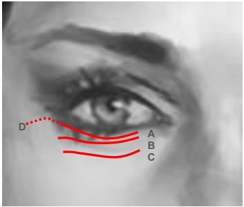
The inferior orbital rim and orbital floor can be accessed via a preseptal transconjunctival approach12; visualisation can be augmented with endoscopy13. Alternatively, a transcutaneous approach can also be made via subciliary, subtarsal, or infra-orbital incisions.
The preseptal transconjunctival approach provides access to the orbital floor and rim. This approach may be combined with lateral canthotomy and cantholysis for extended access to the lateral orbit.
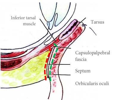
Transcutaneous subciliary, subtarsal, or infraorbital incisions (Figure 26)
The medial orbit can be accessed using a preseptal precaruncular transconjunctival approach 14. Dissection deep to periorbita may be facilitated by using a Freer’s suction elevator. Recent developments in multiportal endoscopic approaches to the skull base have also been reported 15, 16.
Fractures of maxilla and lower midface
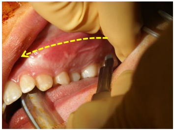
The maxilla and lower midface can be exposed via a sublabial incision positioned approximately 1cm from the labial sulcus to facilitate closure (Figure 27).
Nasoethmoid/Orbital Fractures
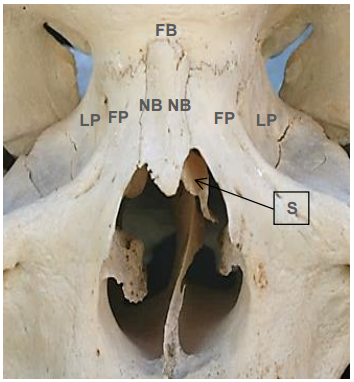
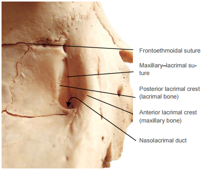
Naso-orbital-ethmoid (NOE) complex fractures generally result from high-energy impact to the glabella or nasal root. Because the lamina papyraceae of the medial orbital walls are extremely thin (Figures 29, 30), these often fracture, resulting in posterior displacement of the nasal root.
This presents a challenge because the fractures can involve the frontal sinus outflow pathways causing sinonasal dysfunction, or cerebrospinal fluid leaks.
NOE fractures are often best visualised with axial CT scans; both bony and soft tissue windows should be reviewed, particularly in the orbital region. Coronal images are helpful to delineate the floor of the anterior cranial fossa.
Surgical access may be technically challenging; accurate reduction and fixation are even more demanding. Fixation of these fractures can be achieved using microplate systems (1.0-1.3mm with 2-3mm screws). Fractures of the orbital floor are can be repaired using bone grafts, titanium mesh, porous polyethylene (PPE) sheets, composite PPE and titanium mesh, or resorbable materials.
The medial canthal ligaments represent the medial extensions of the orbicularis oculi muscles and are of importance in the management of NOE fractures (Figure 31).
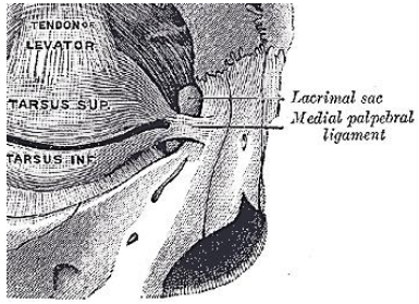
The ligaments split into two primary components prior to their attachments to the anterior and posterior lacrimal crests, with some superior extensions as well (Figure 32). Fractures can displace the medial canthal ligaments resulting in malposition of the globe e.g. telecanthus.
The initial examination of the patient with an orbital wall fracture should include a thorough ophthalmologic examination with particular attention to extraocular muscle function and visual acuity. If either of these is compromised one should consult an ophthalmologist.
Orbital blowout fractures with herniation of orbital soft tissue into the maxillary or ethmoid sinuses and result in decreased orbital volume (enophthalmos) or diplopia due to entrapment of extraocular muscles causing limitation of ocular movements. Diplopia is a common consequence of orbital blowout fractures.
Entrapment of extraocular muscles requires immediate surgical intervention. Delay in reconstituting the periorbita and the entrapped muscle can result in fibrosis of the tissues and permanent diplopia. If a blowout defect is large enough to permit substantial herniation of tissue, hypoglobus and/or enophthalmos can result. In such defects, it is uncommon to have entrapment of extraocular muscles.
In a patient unable to cooperate with the physical examination, forced duction or traction testing (Figure 33) can be used to determine the presence of mechanical restriction of ocular motility.
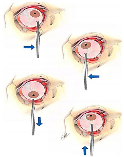
The bulbar conjunctiva is anaesthetised topically and the conjunctiva is grasped near the limbus with toothed forceps. The globe is then moved into the direction in which the mechanical restriction is suspected.
Fractures of the orbital apex or optic canal are not common. In cases of traumatic optic neuropathy, magnetic resonance imaging (MRI) should be employed prior to decompression. (See chapter: Endoscopic transnasal optic nerve decompression)
More than 1/3 of facial fractures affect the nasal complex 17. Examine the external nose and nasal cavity thoroughly. Nasal fractures can be present without causing nasal deformity or airway obstruction.
Epistaxis can be controlled in a number of ways. Initial management may involve pinching the cartilaginous nasal walls against the nasal septum for 15 minutes. If this fails, then the nose is suctioned and oxymetazoline is sprayed into the nose for vasoconstriction and the cartilaginous nose is again pinched for 15 minutes. If this fails, one may elect to insert absorbable or non-absorbable packing in the nasal cavities. Absorbable nasal packing is preferred because it avoids traumatising the fragile nasal mucosa.
It is important to perform an endonasal examination to rule out a septal haematoma as it must be evacuated to prevent a septal abscess or septal necrosis 18. Drainage is performed by incising the septal mucoperichondrium. Silastic splints are placed endonasally to oppose the muco-perichondrial flaps against the septal cartilage.
Nasal fractures are reduced only if the patient has aesthetic or functional complaints. It is the authors’ opinion that radiographic examination is unnecessary in clinically undisplaced nasal fractures (Figure 34). Such patients are simply reminded to avoid contact sports while the nasal bones heal.
Because initial soft tissue swelling may mask a displaced bony fracture, it is prudent to re-examine such a patient once the soft tissue swelling has subsided. Timely reduction of fractures is important as delays make manipulation of fractured bone fragments very challenging and may lead to malunion. Although it may be possible to achieve a closed reduction up to 3 weeks after injury, it is preferable to perform the reduction within 10-14 days after the injury.
Reduction of nasal fractures can often be accomplished via a closed approach. This can typically be performed under local anaesthesia and insertion of nasal pledgets soaked in vasoconstrictor e.g. oxymetazoline or cocaine. An Asch septum-straightening forceps or Boies nasal fracture elevator is typically used to reduce the fracture. The Boies elevator is inserted endonasally on the depressed side first with the dominant hand and the surgeon’s non-dominant hand is placed on the nose for stability. The fracture is then reduced into place. The same process is performed on the contralateral side to reduce the fractured fragment. Adhesive strips and a splint are placed over the nasal dorsum, and are removed after 5-10 days. In patients with extensive fractures, open septal fractures, or persistent deformities following closed reduction, open reduction can be considered.
Le Fort fractures typically occur in conjunction with other midface fractures. If only one side of the face is involved, it is termed a “hemi” Le Fort fracture. Le Fort I fractures traverse the maxillary alveolus through the pyriform sinus and may involve the pterygoid plates. Le Fort II fractures extend from the maxilla into the orbit in a pyramidal configuration. Le Fort III fractures are complete craniofacial dissociation.
Although these fracture patterns are useful descriptors to communicate between providers, craniomaxillofacial surgeons should describe each fracture in detail on each side of the face, also including fractures of the frontal bone and mandible.
Miniplate systems (1.5-2.0 mm with 4-6 mm screws) are ideal for fixation of lower midface fractures.
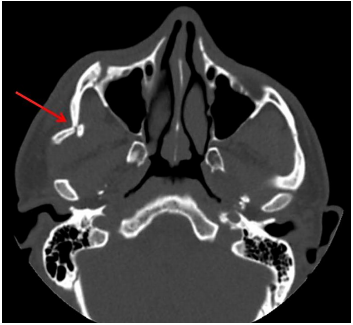
Zygomaticomaxillary complex fractures involve the zygomatic arch, in addition to disruption of its articulations with the frontal, sphenoid, maxillary and temporal bones. Axial CT scans readily identify such fractures (Figure 36).
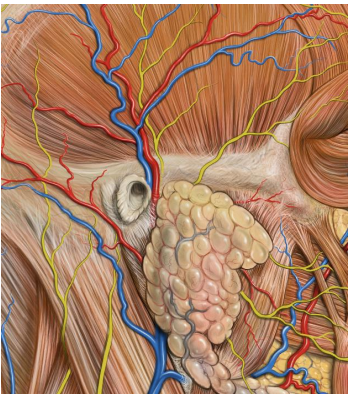
These fractures are reduced using approaches highlighted earlier. Plating can be challenging in this region because of the proximity of the frontal branch of the facial nerve to the zygoma (Figure 37).
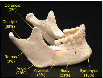
Mandibular fractures are the second most prevalent type of facial fracture 19 and most commonly involve the condylar region, followed by the body and angle (Figure 38).
Fractures can be described as greenstick, simple, or compound. Greenstick fractures entail injury to only one surface of the bone, and are common in the paediatric population because of the flexibility of the young skeleton. A simple fracture is a fracture not involving the intraoral mucosa. Compound fractures, also known as open fractures, involve exposure of bone via violations in soft tissue. Comminuted fractures have multiple fragments of bone disassociated from one another.
Open reduction and internal fixation of mandible fractures can be straightforward as parasymphyseal fractures, or very challenging as in subcondylar fractures. Consequently complication rates following mandible fracture repair are significant. A recent series of >500 mandible fractures revealed an overall complication rate of 26% with hardware failure being the most common 20. The authors found that antibiotic use did not decrease the infection rates, but that patients with systemic medical conditions and smokers had higher risks of postoperative complications.
Substantial variations exist in the treatment of mandibular fractures around the globe. Otolaryngologists, plastic surgeons, and oromaxillofacial surgeons all operate on the mandible and come from different training backgrounds, which likely accounts for some of the variations in treatment. Nonetheless there are several aspects of treatment that ought to be practiced universally.
The 1st step is to determine if the patient’s dentition is adequate for placement of maxillo-mandibular fixation (MMF). In the edentulous patient, open reduction and internal fixation (ORIF) is necessary for repair of displaced mandible fractures, whereas in the dentate mandible, if teeth are present on both the mesial and distal aspects of the fracture line, ORIF or closed reduction are both reasonable options.
Closed reduction of is an option for no or minimally-displaced simple fractures that can undergo MMF. Any comorbid condition that may preclude ORIF e.g. a medically unstable patient, is also an indication for closed reduction. Closed reduction is accomplished with placement of arch bars and the application of dental wiring to achieve MMF. The MMF is released 6 weeks later and fracture stability is assessed. If the patient remains in occlusion, then he/she should be placed on a soft diet and arch bars should be removed 2 weeks later. Postoperative panorex examination can be helpful to assess bony alignment and occlusion.
This requires reduction of multiple small fragments and is often best managed via a transcutaneous approach.

These can be accessed via retromandibular (rhytidectomy) approaches (Figure 41). While it permits excellent access to the posterior mandible, it does carry a great risk of damage to the facial nerve. Even slight traction on the facial nerve can cause postoperative dysfunction which can be disturbing for patients. For this reason, endoscopic approaches to the subcondylar region, although technically challenging, are also be used.
Treatment can be complex, and can entail observation, closed reduction, or ORIF based on the extent of injury 5.
AO Surgery: Guide to Maxillofacial Trauma (Highly recommended)
Prabhat K. Bhama, M.D., M.P.H.
Staff Surgeon
Department of Otolaryngology
Alaska Native Tribal Health Consortium
Anchorage, Alaska, USA
pbhama@gmail.com
Mack L. Cheney, M.D.
Chair, Office of Global Surgery
Professor, Division of Facial Plastic and Reconstructive Surgery
Department of Otolaryngology
Harvard Medical School/Massachusetts Eye and Ear Infirmary
Boston, Massachusetts, USA
mack_cheney@meei.harvard.edu
Johan Fagan MBChB, FCORL, MMed
Professor and Chairman
Division of Otolaryngology
University of Cape Town
Cape Town
South Africa
johannes.fagan@uct.ac.za