Selective neck dissection (SND) entails removal of cervical lymph nodes only from selected levels of the neck. It is generally done as an elective neck dissection (END) i.e. in the absence of clinically apparent cervical metastases when the risk of having occult cervical nodal metastases is thought to exceed 15-20%; or for very limited nodal metastases. It may be technically more demanding than a modified neck dissection (MND) due to poorer exposure, and requires a sound knowledge of the 3-dimensional anatomy of the neck.
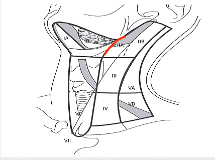
The neck is conventionally divided into 6 levels; Level VII is in the superior mediastinum (Figure 1).
Level I is bound by the body of the mandible above, the stylohyoid muscle posteriorly, and the anterior belly of the contralateral digastric muscle anteriorly. The revised classification (Figure 1) uses the posterior margin of the submandibular gland as the boundary between Levels I and II as it is clearly identified on ultrasound, CT, or MRI. Level I is subdivided into Level Ia, (submental triangle) which is bound by the anterior bellies of the digastric muscles and the hyoid bone, and Level Ib (submandibular triangle).
Level II extends between the skull base and hyoid bone. The posterior border of the sternocleidomastoid defines its posterior border. The stylohyoid muscle (alternately the posterior edge of the submandibular gland) defines its anterior border. The accessory nerve (XIn) traverses Level II obliquely and subdivides it into Level IIa (anterior to XIn) and Level IIb (behind XIn).
Level III is located between the hyoid bone and the inferior border of the cricoid cartilage. The sternohyoid muscle marks its anterior limit and the posterior border of the sternocleidomastoid its posterior border.
Level IV is located between the inferior border of the cricoid cartilage and the clavicle. The anterior boundary is the sternohyoid muscle, and the posterior border is the posterior border of sternocleidomastoid.
Level V is bound anteriorly by the posterior border of the sternocleidomastoid, and posteriorly by the trapezius muscle. It extends from the mastoid tip to the clavicle, and is subdivided by a horizontal line drawn from the inferior border of the cricoid cartilage into Level Va superiorly, and Level Vb inferiorly.
Level VI is the anterior, or central, compartment of the neck. It is bound laterally by the carotid arteries, superiorly by the hyoid bone, and inferiorly by the suprasternal notch.
Neck dissection operations are classified according to cervical lymphatic levels that are resected (Figures 1, 2).
Selective neck dissections: Commonly performed SNDs are illustrated in Figure 2, and include lateral, posterolateral, supraomohyoid, anterolateral and central SND.
Lateral ND (Levels II-IV) is commonly employed for cancers of oropharynx, hypopharynx and larynx; Posterolateral ND (Levels II-V) is used for skin cancers posterior to the ear; Supraomohyoid ND (Levels I-III) is typically indicated for
cancers of the oral cavity other than cancers of the anterior tongue and floor of mouth when an Anterolateral ND (Level I-IV) is preferred so as to encompass skip metastases to level IV. Central neck dissection encompasses only Level VI and is used with thyroid cancer (Figure 1).
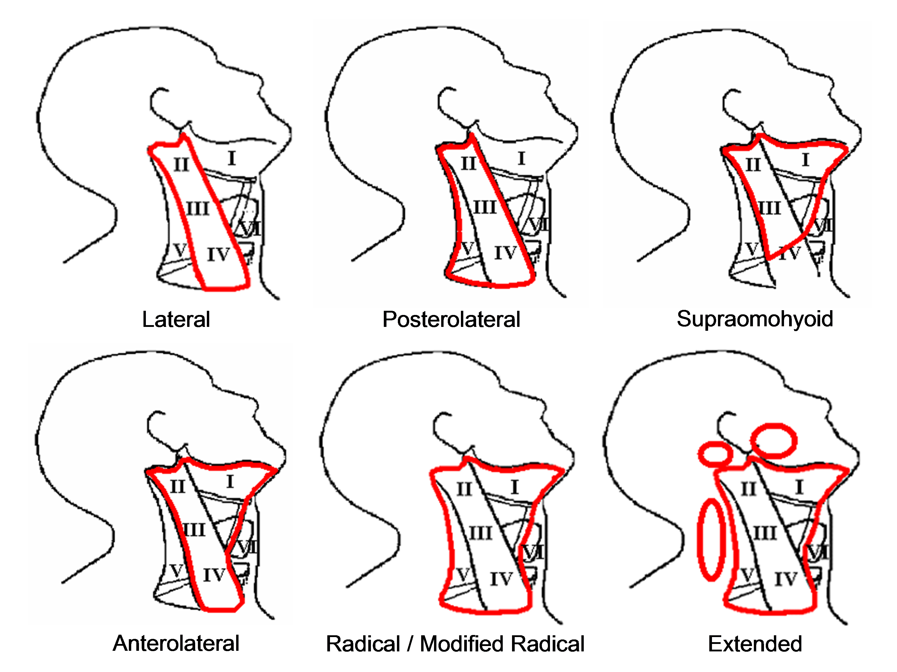
Comprehensive or therapeutic neck dissection involves surgical clearance of
Levels 1-V and may either be a radical (RND) or modified (MND) neck dissection. RND includes resection of sternocleidomastoid muscle (SCM), accessory nerve (XIn) and internal jugular vein (IJV). MND preserves SCM and/or XIn and/or IJV.
Extended neck dissection includes resection of additional lymphatic groups (parotid, occipital, Level VI, mediastinal, retropharyngeal) or non-lymphatic structures (skin, muscle, nerve, blood vessels etc.) not usually included in a comprehensive neck dissection.
It has been proposed that neck dissections be more logically and precisely described and classified by naming the structures and the nodal levels that have been resected. (Ferlito A, Robbins KT, Shah JP, et al. Proposal for a rational classification of neck dissections. Head Neck 2011 Mar; 33(3): 445-50)
The operation is done under general anaesthesia without muscle relaxation as eliciting muscle contraction on mechanical or electrical stimulation of the marginal mandibular, hypoglossal (XIIn) and acces-sory nerves assists with locating and preserving these nerves. It is a clean operation and antibiotics are therefore not required unless the upper aerodigestive tract is entered. With an experienced surgeon, blood transfusion is rarely required.
The patient is placed in a supine position with the neck extended and head turned to the opposite side. Surgical draping must permit monitoring for movement of the lower lip with irritation of the marginal mandibular nerve, and must provide access to the clavicle inferiorly, the trapezius muscle posteriorly, the tip of the earlobe superiorly and the midline of the neck anteriorly. The drapes are sutured to the skin.
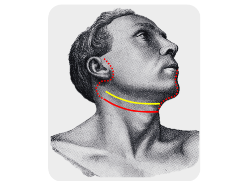
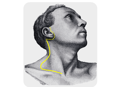
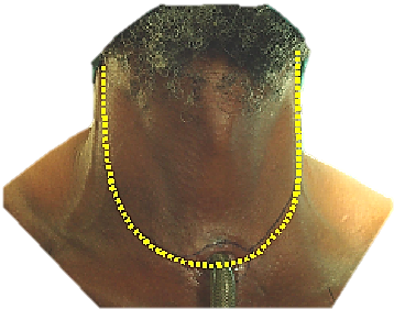
Incisions should take into consideration access that may be required to resect the primary tumour, cosmetic factors, and the blood supply to the flaps. A transverse skin crease incision is placed more inferiorly than with a MND so as to avoid a vertical skin incision and to facilitate dissection of levels III and IV (Figure 3). The transverse skin incision can be extended across to the opposite side with bilateral SND, or can be extended superiorly to split the lower lip in the midline to gain access to the oral cavity, or preauricularly for a parotidectomy (Figure 3). Figure 4 demonstrates the hockey stick incision. The hockey-stick incision may be extended into a preauricular skin crease and is particularly useful for combined posterolateral ND and parotidectomy. Care has to be taken in patients who have been previously irradiated as the posteroinferior corner of the flap has a tenuous blood supply and may slough and have to heal by secondary intention.
Total laryngectomy with Lateral ND can be accessed via a wide apron flap (Figure 5).
The detailed step-by-step description of neck dissection that follows refers to a right-sided Supraomohyoid ND (Levels I-III). For a Lateral ND, simply skip dissecttion of Level 1 and extend the nodal resection inferiorly beyond the level of the omohyoid muscle, either by retracting or dividing the muscle for access.
The neck is opened via a horizontal incision placed in a skin crease just below the level of the hyoid bone. The incision is made through skin, subcutaneous fat, and platysma muscle. Identify the external jugular vein and greater auricular nerve overlying the SCM (Figure 6).
Next the superior skin flap is elevated with cautery in a subplatysmal plane until the submandibular salivary gland is identified. The surgeon then uses electrocautery or a scalpel to raise an inferiorly based sub-platysmal flap, exposing the neck as follows: anteriorly up to the omohyoid muscle (the posterior margin of which corresponds to the anterior margin of the supraomohyoid or anterolateral neck dissections) and inferiorly, the lateral surface of the SCM almost to the clavicle The only structure to look out for and not injure during this step of the dissection is the external jugular vein which lies on the lateral surface of the sternomastoid muscle.
The recommended subsequent operative steps of a supraomohyoid neck dissecttion are illustrated in Figure 7.
The surgeon resects fat and lymph nodes from the submental triangle (Level Ia). The skin is elevated in a subplatysmal plane up to the opposite anterior belly of digastric muscle, looking out for the anterior jugular veins. The contents of the sub-mental triangle are resected with electrocautery up to the hyoid bone. The deep plane of dissection is the mylohyoid muscle (Figures 8, 9).
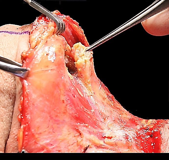
The surgeon next addresses Level Ib of the neck. The fascia (capsule) overlying the submandibular gland is incised midway over the gland and is dissected from the gland in a superior direction in a subcapsular plane so as to avoid injury to the marginal mandibular nerve (Figure 10). Using this technique the marginal mandibular nerve does not need to be routinely identified; the assistant however watches for twitching of the lower lip as this indicates proximity to the nerve. The marginal mandibular nerve crosses the facial artery and vein (Figure 11).The facial artery and vein are identified by blunt dissection with a fine haemostat (Figure 11).
Next attention is directed to the fat and lymph nodes tucked anteriorly between the anterior belly of digastric and mylohyoid muscle (Figure 11). These nodes are especially important to resect with malignancies of the anterior floor of mouth. To resect these nodes one retracts the anterior belly of digastric anteriorly and delivers the tissue using electrocautery dissection with the deep dissection plane being on the mylohoid muscle (Figures 12, 13).
Other than the nerve to mylohoid and vessels that pierce the muscle and need to be cauterized or ligated, there are no significant structures until the dissection reaches the posterior free margin of the mylohyoid muscle.
Next attention is directed at the region of the facial artery and vein. The surgeon palpates around the facial vessels for facial lymph nodes; if present, they are dissected
free using fine haemostats, taking care not to traumatise the marginal mandibular nerve. The facial artery and vein are then ligated and divided close to the submandibular gland so as not to injure the marginal mandibular nerve (Figure 12). This frees up the gland superiorly, which can then be reflected away from the mandible (Figure 13).
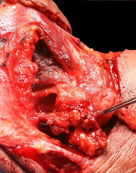
Next the surgeon addresses the lingual nerve, submandibular duct, and XIIn. The mylohyoid muscle is retracted anteriorly with a right-angled retractor. The clearly defined interfascial dissection plane between the deep aspect of the submandibular gland and the fascia covering the XIIn is opened with finger dissection, taking care not to tear the thin-walled veins accompanying XIIn. The XIIn is now visible in the floor of the submandibular triangle (Figure 14). Inferior traction on the gland brings the lingual nerve and the submandibular duct into view (Figure 14).
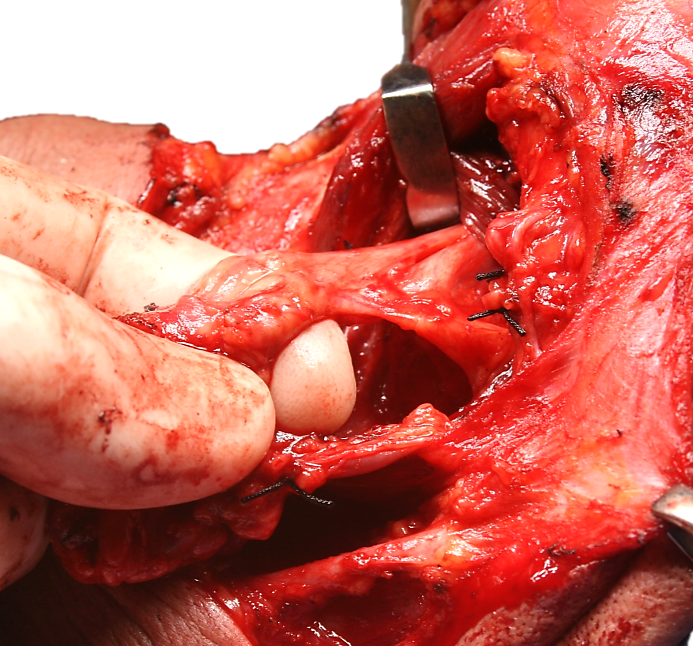
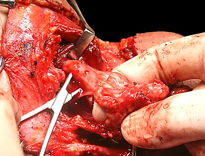
The submandibular duct is separated from the lingual nerve, ligated and divided (Figures 15, 16). The submandibular ganglion, suspended from the lingual nerve, is clamped, divided and ligated, taking care not to cross-clamp the lingual nerve (Figure 16).
The facial artery is divided and ligated just above the posterior belly of digastric (Figure 17).

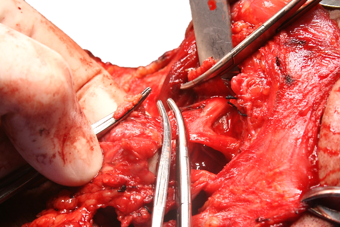
Note: A surgical variation of the above technique is to preserve the facial artery by dividing and ligating the 1-5 small branches that enter the submandibular gland. This is usually simple to do, it reduces the risk of injury to the marginal mandibular nerve, and permits the use of a buccinator flap based on the facial artery (Figure 18).
This step entails identifying the XIIn in Level IIa, and tracing the XIIn posteriorly to where it leads the surgeon directly to the internal jugular vein (IJV).
Divide the external jugular vein (Figure 19). This is a key step with SND as it improves access to Levels IIa and IIb. The greater auricular nerve is preserved.
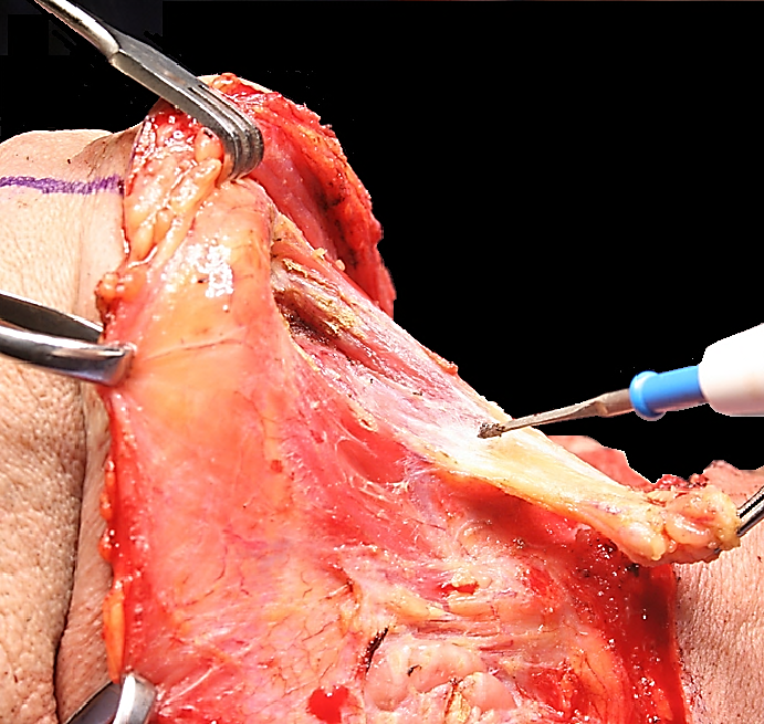
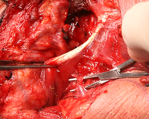
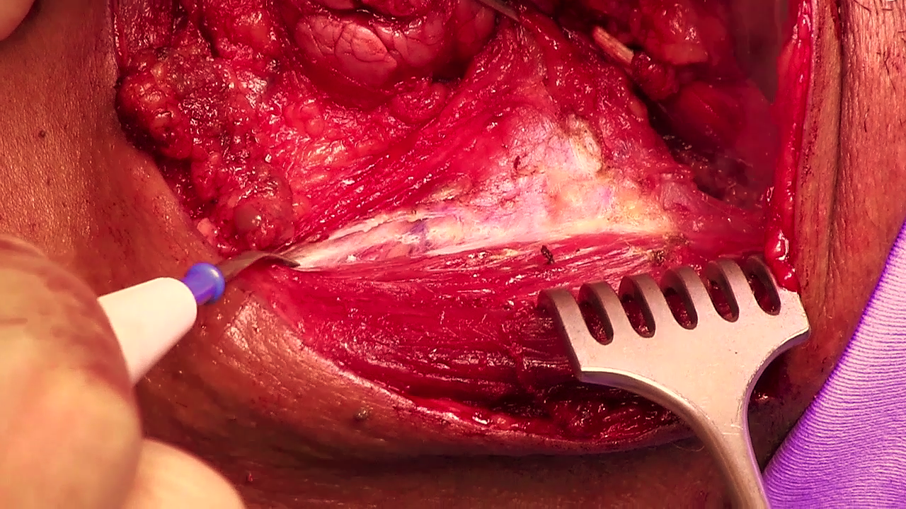
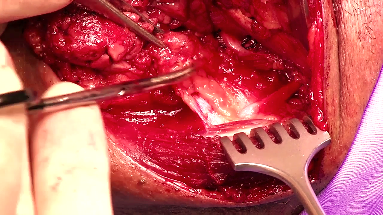
Divide the fascia along the lateral aspect of the posterior belly of digastric (Figure 20). This step is the key to facilitating subsequent exposure of the IJV and XIn. Expose the posterior belly of digastric along its entire length, taking care not to wander above the muscle as this might jeopardise the facial nerve. No significant structures cross the posterior belly other than the facial vein.
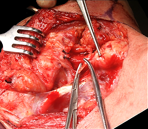
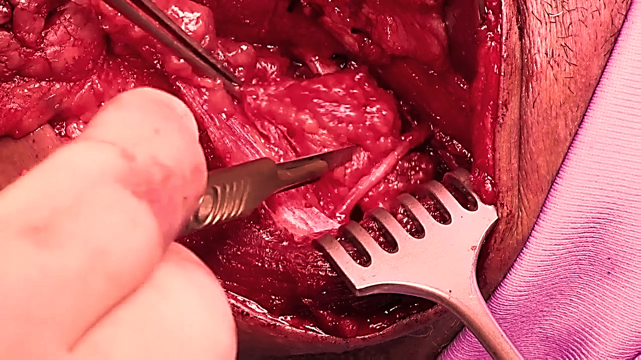
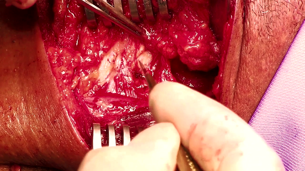
Next identify the XIIn below the greater cornu of the hyoid bone anterior to where it crosses the external carotid artery. It is generally more superficial than expected, and is located just deep to the veins that cross the nerve. Carefully dissect along the nerve in a posterior direction and divide all the veins crossing the nerve to expose the full length of XIIn (Figure 21).
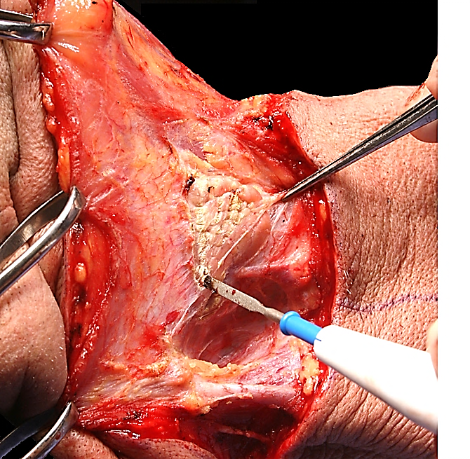
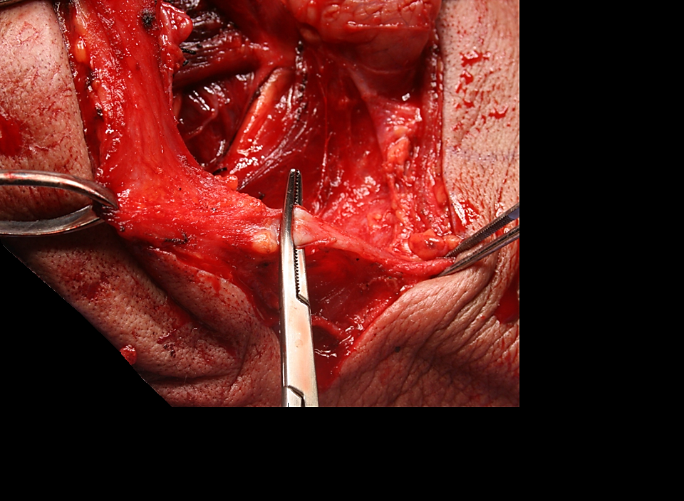
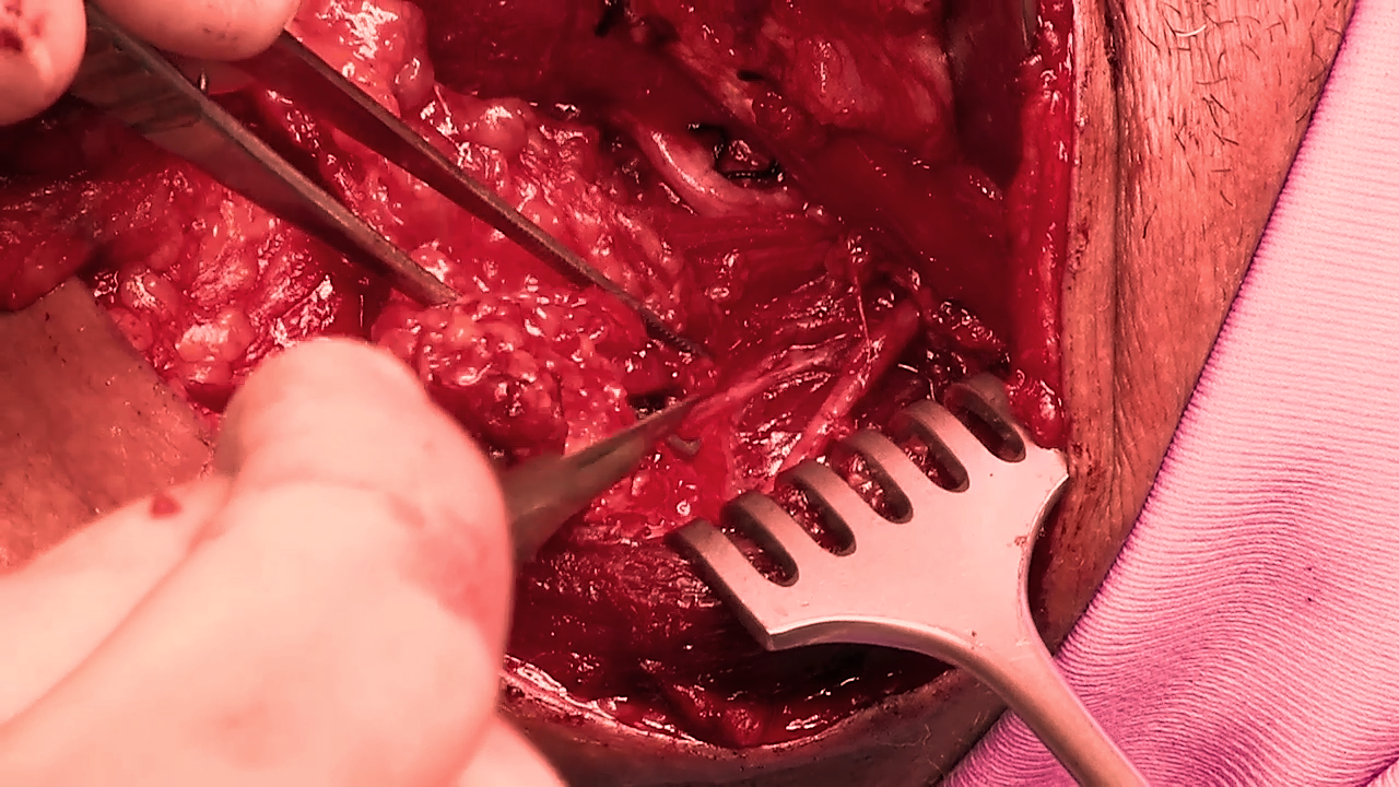
After the nerve has crossed posterior to the external carotid artery, identify the SCM branch of the occipital artery that tethers the XIIn (Figure 22).
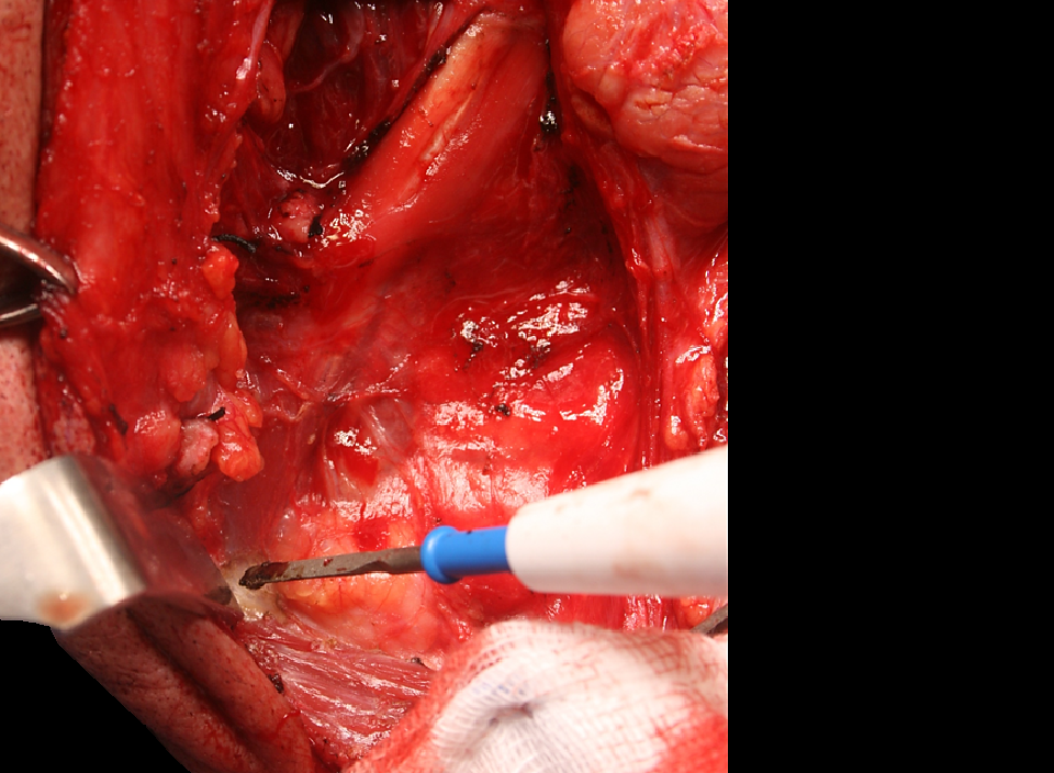
Dividing this artery releases the XIIn (Figure 23). The nerve then courses vertically along the anterior surface of the IJV and hence leads the surgeon directly to the IJV (Figure 23).
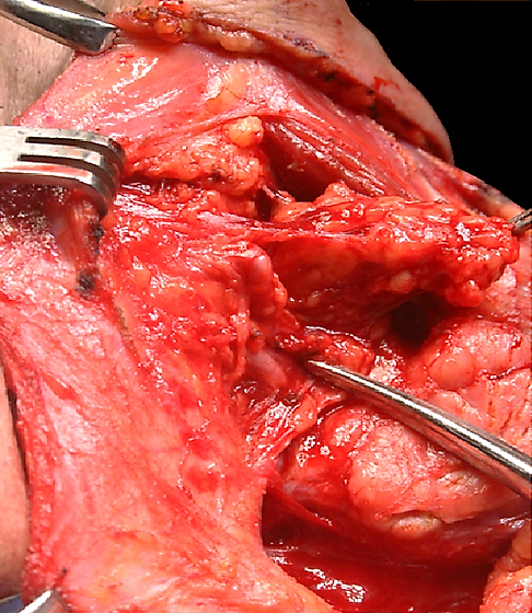
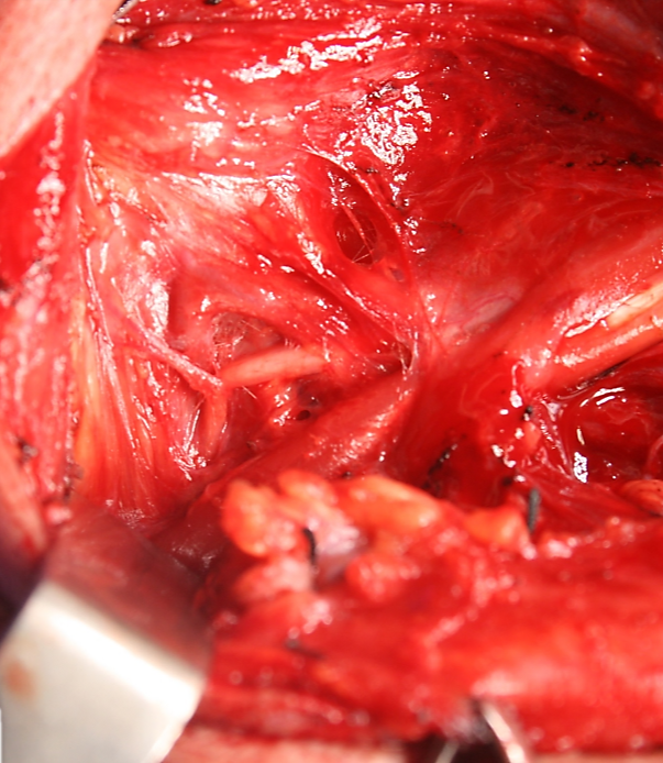
Using dissecting scissors or a haemostat to part the fatty tissue behind the IJV in Level II, the surgeon next identifies the XIn which may course lateral (commonly), medial (uncommonly) or through (very rarely) the IJV. The nerve is often first located by noting movement of the shoulder due to mechanical stimulation of the nerve (Figure 23).
Dissect with a scalpel or electrocautery along the omohyoid and strip the fatty tissue in the anterior parts of Levels II and III from the underlying infrahyoid strap muscles in a posterior direction towards the carotid sheath. Divide the epimysium along the anterior border of the SCM using electrocautery or a scalpel (Figure 24). This exposes structures deep to the SCM i.e. the remainder of Levels II and III of the neck and the lateral surface of the omohyoid muscle as it crosses the internal jugular vein. A number of small vessels entering the muscle are encountered and cauterized. The dissection is carried posteriorly along the deep aspect of the muscle in a subepimysial plane up to the posterior edge of the SCM.
Attention is now directed to clearing Level IIb which is located posterior to the IJV and deep to SCM. Opinions differ as to whether Level IIb (posterior to XIn) needs to be routinely dissected so as to minimise trauma to the XIn.
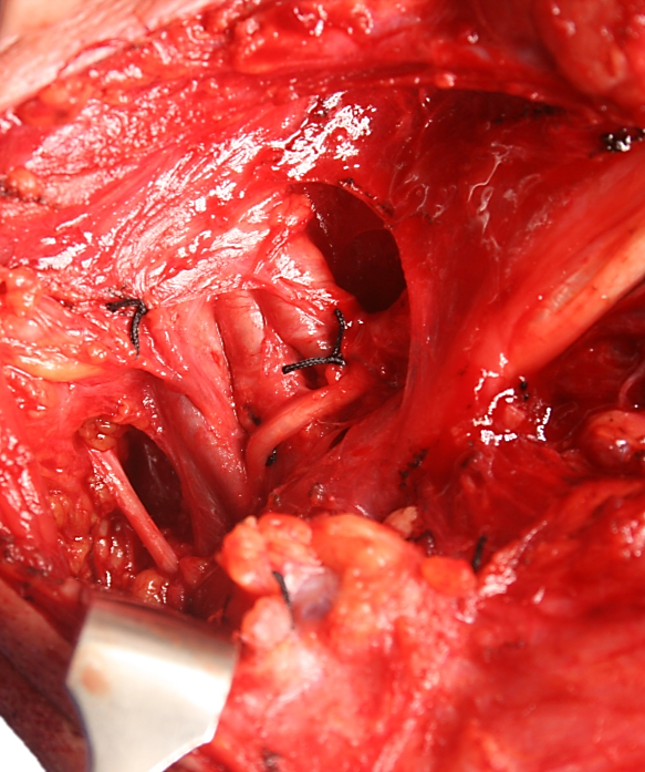
The upper part of the SCM is retracted posteriorly to expose Level IIb. With a haemostat, create a tunnel immediately posterior to the IJV down to the prevertebral muscles (Figure 23). This manoeuver speeds up the subsequent dissection of Level IIb by clearly delineating the posterior wall of the IJV. The transverse process of the C1 vertebra can be palpated immediately posterior to the XIn and IJV and serves as an additional landmark for the position of these structures in difficult surgical cases.
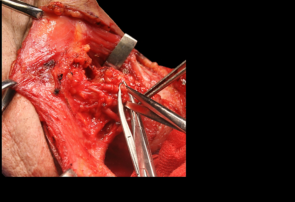
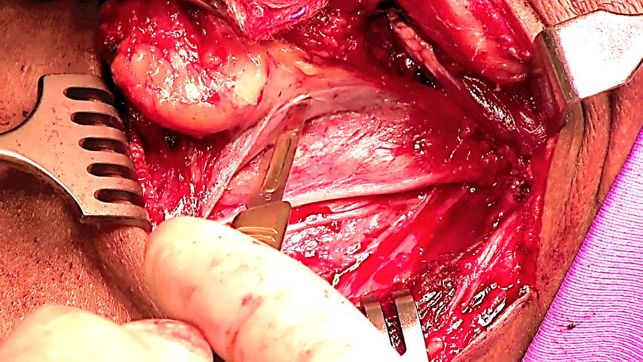
In order to resect Level IIb, identify the XIn in Level IIb, and atraumatically dissect it free from the surrounding fat with sharp and blunt dissection up to where it enters the SCM (Figure 23, 25-27).
Using a scalpel (due to proximity of XIn), or blunt dissection with a haemostat, proceed to mobilise Level IIb starting posterosuperiorly, with the assistant retracting the fatty tissue in an anterior direction. The occipital artery passes across to the top of Level IIb; its branches may need to be cauterized should they be severed while dissecting the superior part of Level IIb. Cut down onto the deep muscles of the neck which are seen to course in a posteroinferior direction. Once the fat of Level IIb has been fully mobilized from the underlying muscles, pass it anteriorly underneath the XIn (Figure 26, 27).
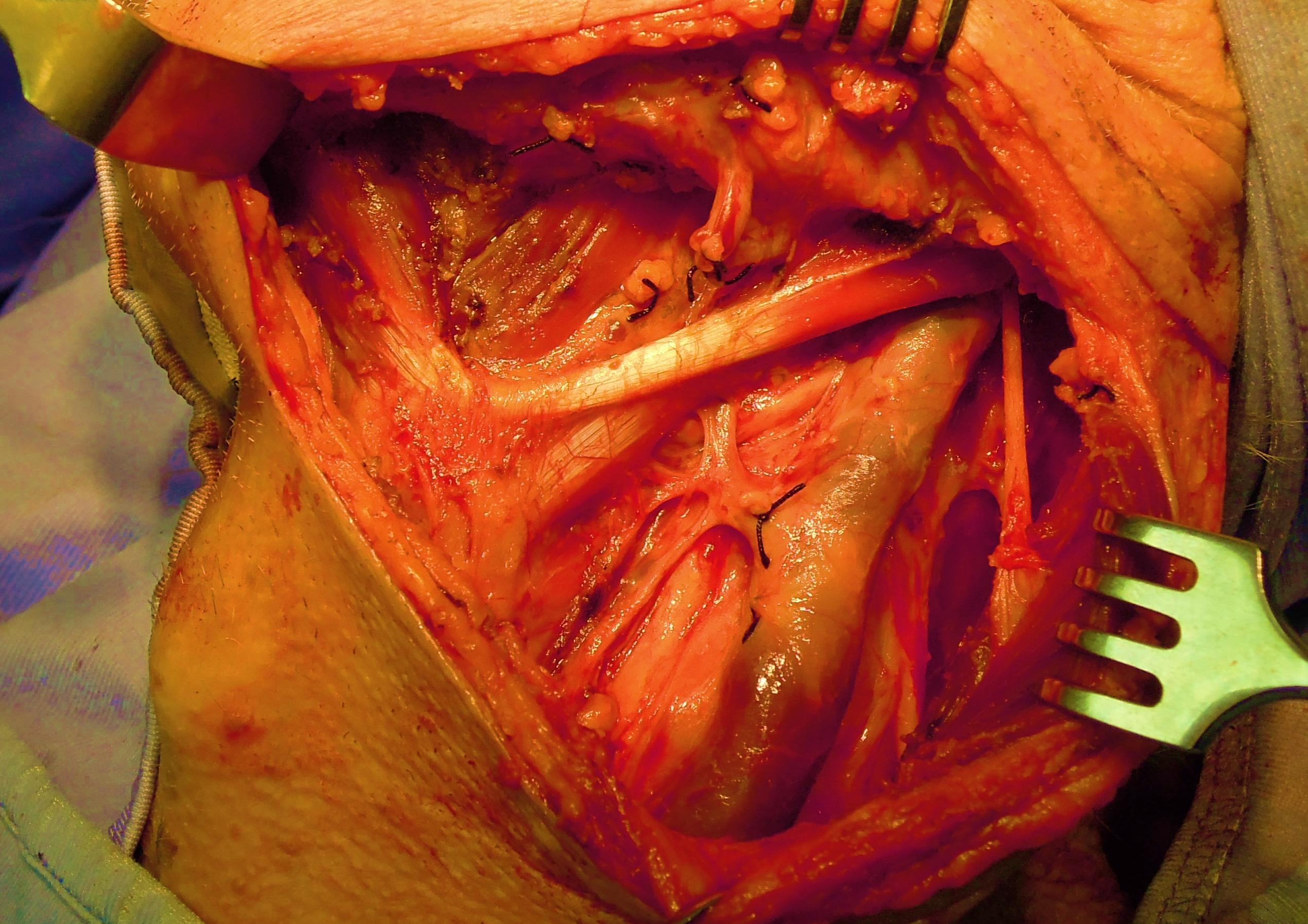
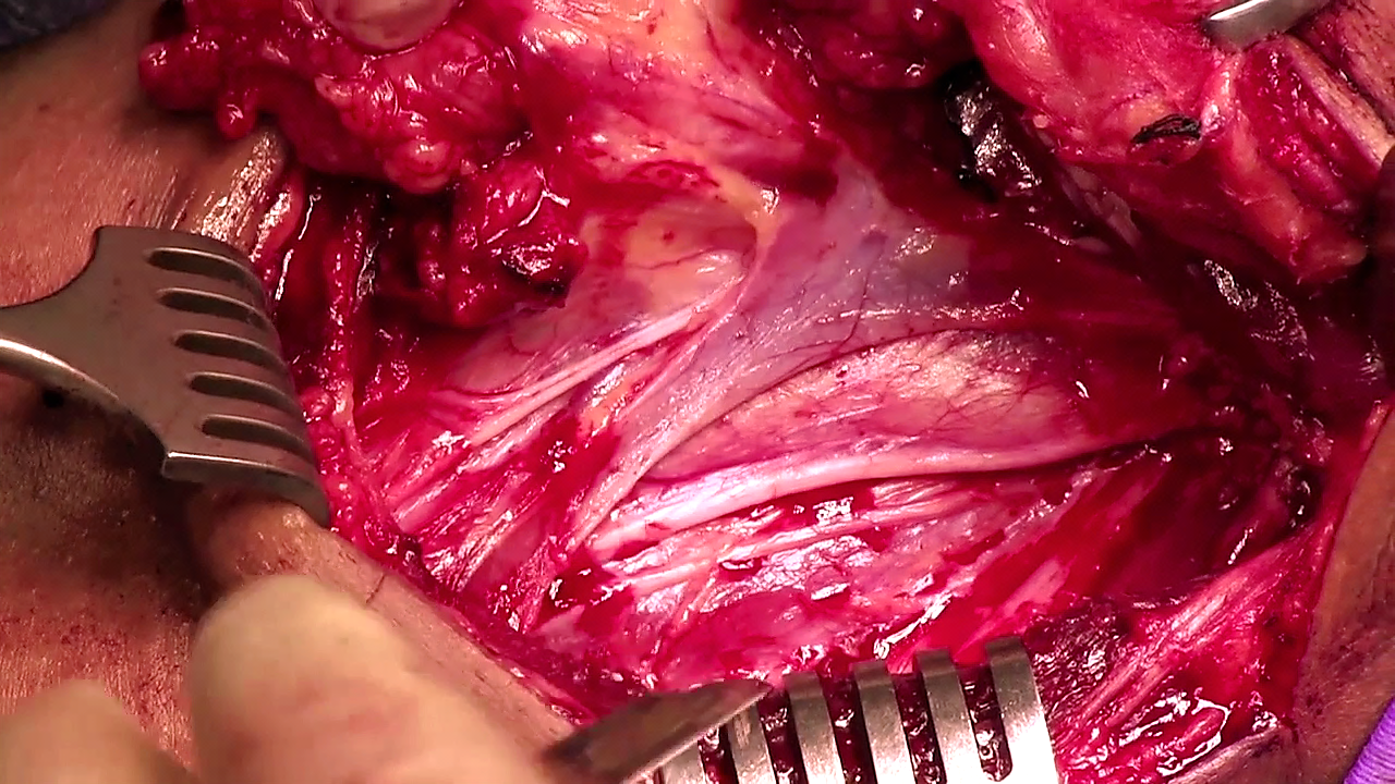
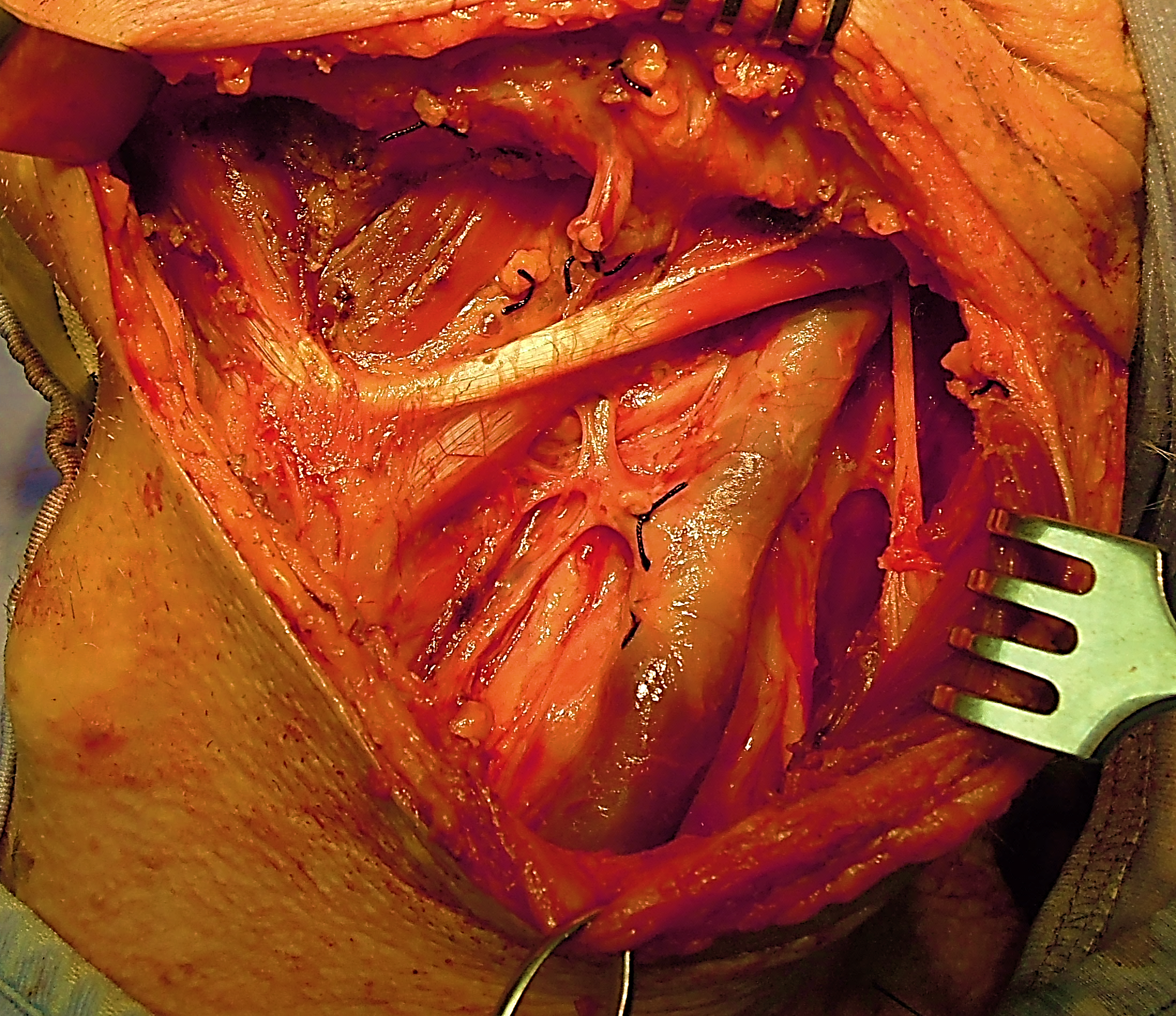
To resect Levels II and III, extend the incision along the posterior edge of the deep aspect of SCM inferiorly through the fatty tissue of Level 3. With the assistant(s) retracting the SCM posteriorly and the fat of levels II and III anteriorly with sharp-toothed rake retractors, dissect the fatty tissue of Levels II and III in an anterograde direction. The deep dissection plane is the muscle the floor of neck between the branches of the cervical plexus which need to be identified and preserved (Figure 28). The phrenic nerve and brachial plexus are not seen in this dissection, but are relevant if Level 4 is dissected. Continue the anterograde dissection with a scalpel or scissors until the ansa cervicalis, and the carotid sheath containing the common and internal carotid arteries, Xn and IJV are sequentially exposed (Figure 29).
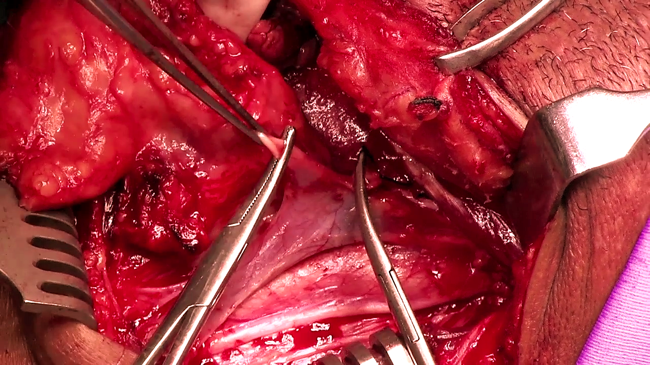
The carotid sheath is incised along the full course of the vagus nerve, and the neck dissection specimen is stripped off the IJV while dissecting inside the carotid sheath. The ansa cervicalis, which courses either deep or superficial to the IJV may be preserved (Figures 29, 30).
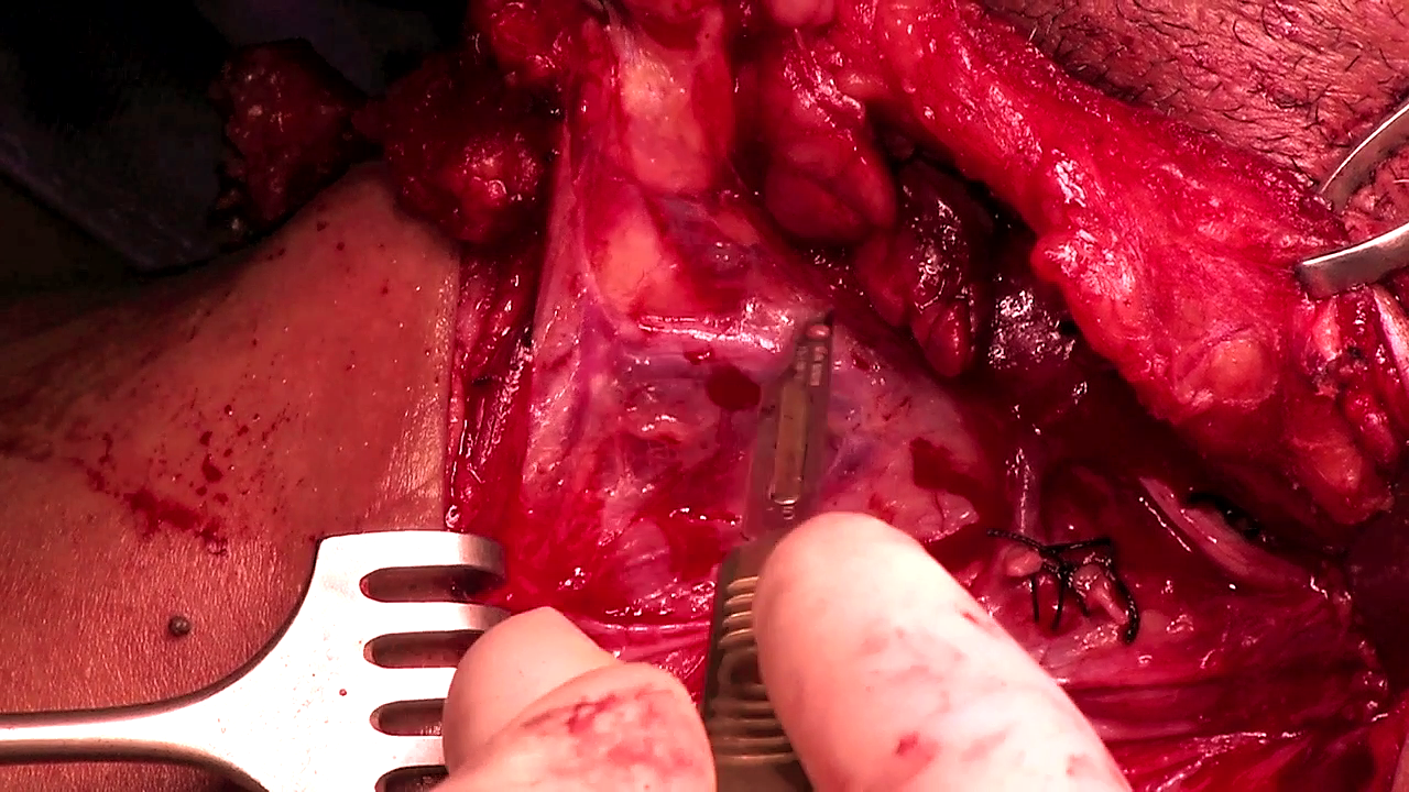
Continue stripping the fat and lymphatics around the anterior aspect of IJV until the common carotid artery is again reached. Divide and ligate tributaries of the IJV with silk ties (Figure 31).
Inferiorly the fatty tissue at the junction of Levels III and IV is divided at the level of the omohyoid (supraomohyoid neck dissection). Identify and preserve the superior thyroid artery where it originates from the external carotid artery (Figure 32).
With a Lateral ND Level IV is resected by applying traction to the fatty tissue deep to the omohyoid in a cephalad direction while dissecting it from Level IV with a scalpel; the transverse cervical vessels may be encountered and need to be ligated; finger dissection may be used to establish a dissection plane between the fat of Level IV and the brachial plexus and phrenic nerve; be vigilant for a chylous leak as the thoracic duct (left neck) or right lymphatic duct may be transected.
The final step is to complete stripping the neck dissection specimen off the infrahyoid strap muscles taking care not to injure the XIIn and its accompanying veins superiorly, and to deliver the neck dissection specimen (Figure 33).
The neck is irrigated with warm water, the anaesthetist is asked to do a valsalva maneuver so as to elicit unsecured bleeding vessels and chyle leakage, and a 5mm suction drain is inserted. The neck is closed in layers with continuous vicryl to platysma and sutures/staples to skin.
The drain is maintained on continuous suction e.g. low pressure wall suction, until the drainage volume is <50ml /24hrs.
Robbins KT, Shaha AR, Medina JE, et al. Consensus statement on the classification and terminology of neck dissection. Arch Otolaryngol Head Neck Surg 2008;134: 536–8.
Ferlito A, Robbins KT, Shah JP, et al Proposal for a rational classification of neck dissections. Head Neck. 2011 Mar;33(3):445-50.
Harris T, Doolarkhan Z, Fagan JJ. Timing of removal of neck drains following head and neck surgery. Ear Nose Throat J. 2011 Apr;90(4):186-9.
Johan Fagan MBChB, FCORL, MMed
Professor and Chairman
Division of Otolaryngology
University of Cape Town
Cape Town
South Africa
johannes.fagan@uct.ac.za