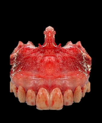
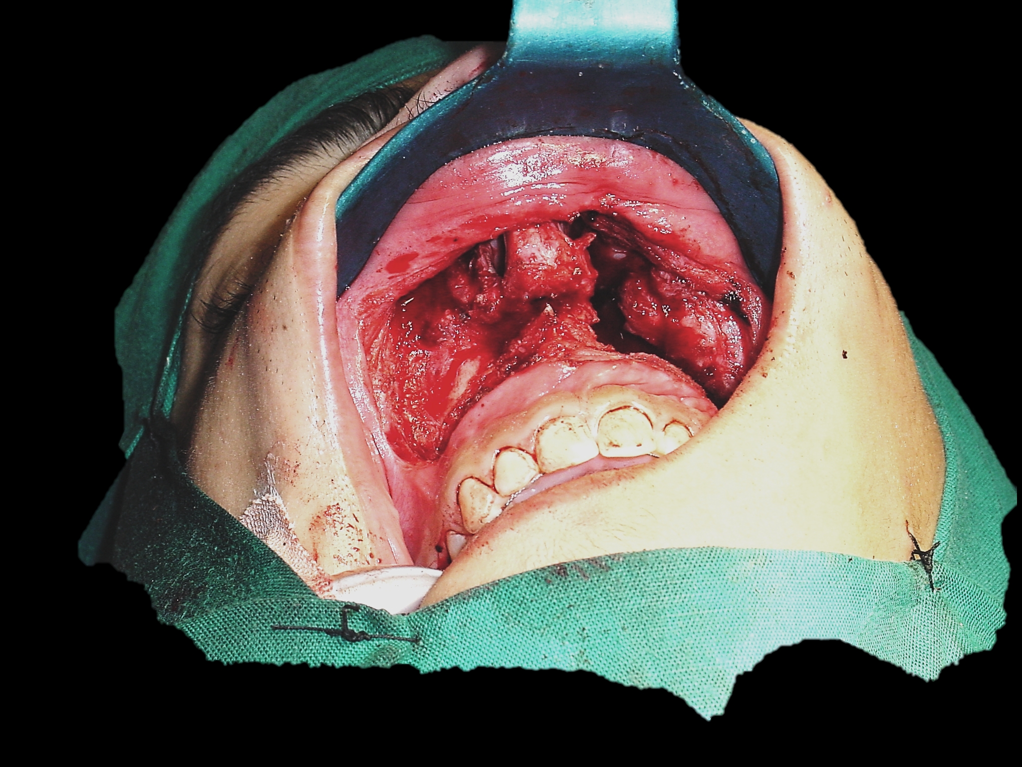
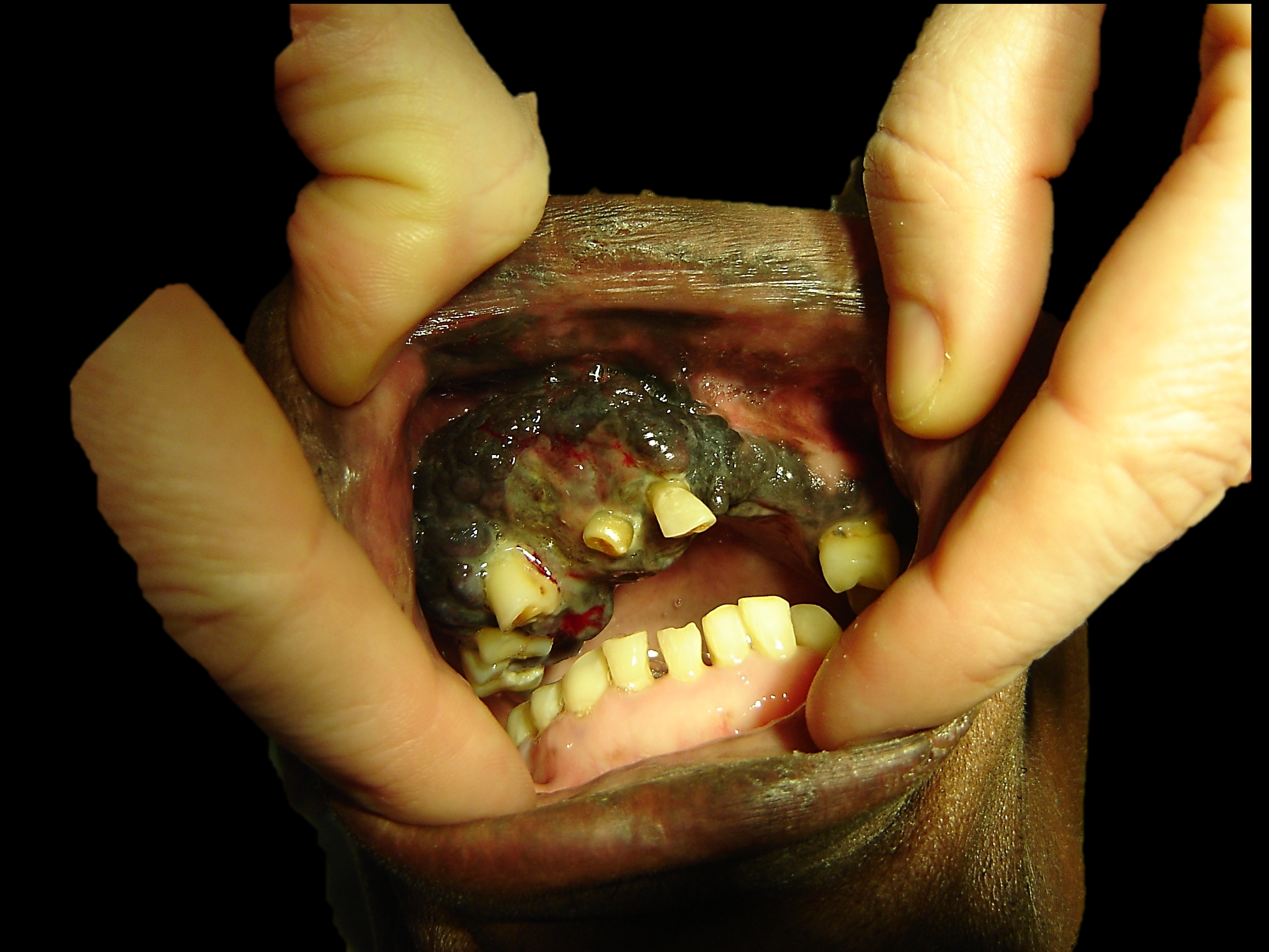
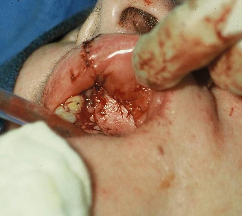
Tumours of the hard palate and superior alveolus may be resected by inferior maxillectomy (Figure 1). A Le Fort 1 osteotomy may also be used as an approach to e.g. angiofibromas and the nasopharynx.
A sound understanding of the 3-dimensional anatomy of the maxilla and the surrounding structures is essential to do the operation safely. Hence the detailed description of the relevant surgical anatomy that follows.
Figure 1: Bilateral inferior maxillectomy
A sound understanding of the 3-dimensional anatomy of the maxilla and the surrounding structures is essential to do the operation safely. Hence the detailed description of the relevant surgical anatomy that follows.
Surgical Anatomy
Bony anatomy
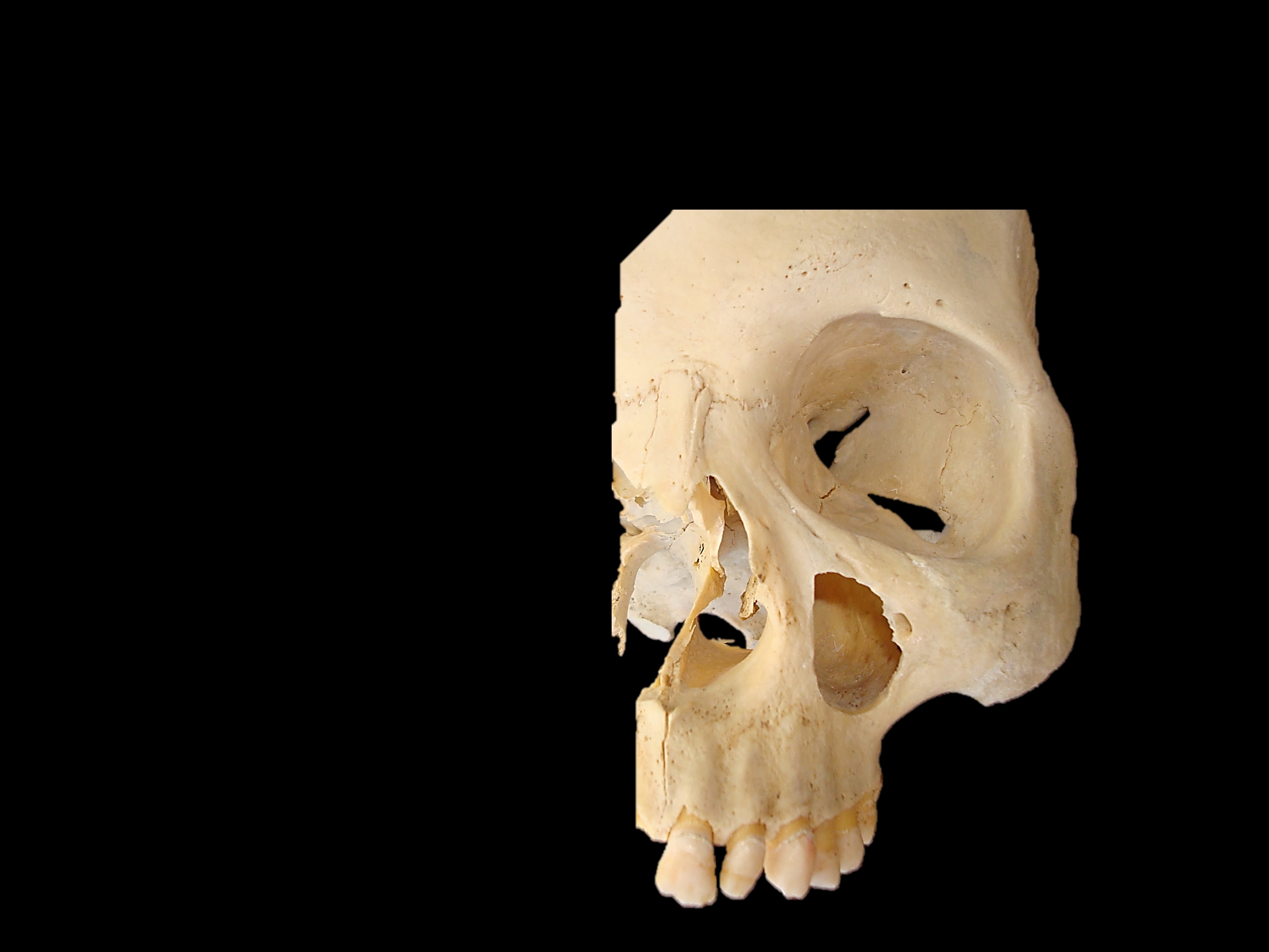
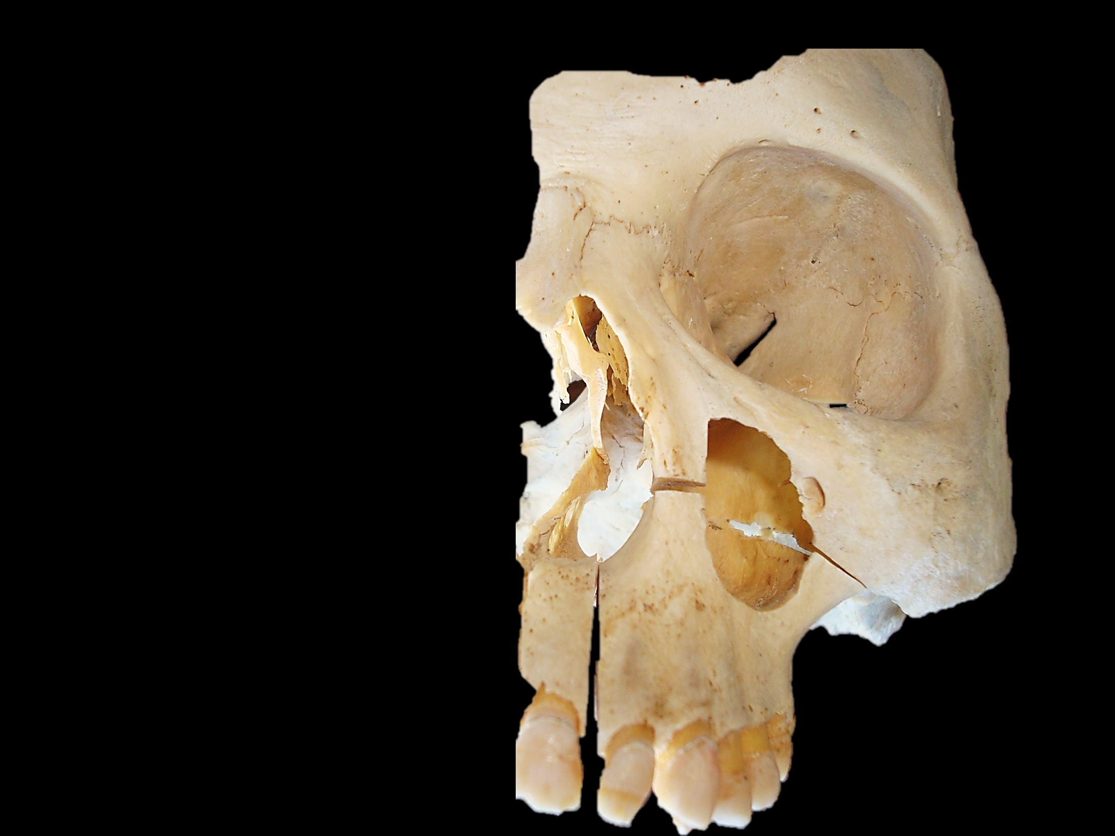
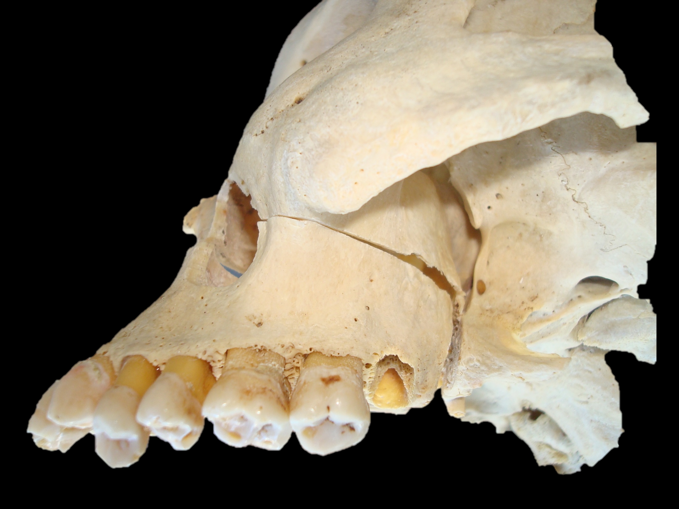
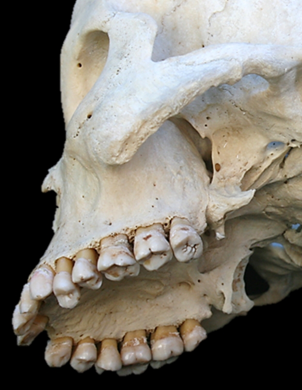
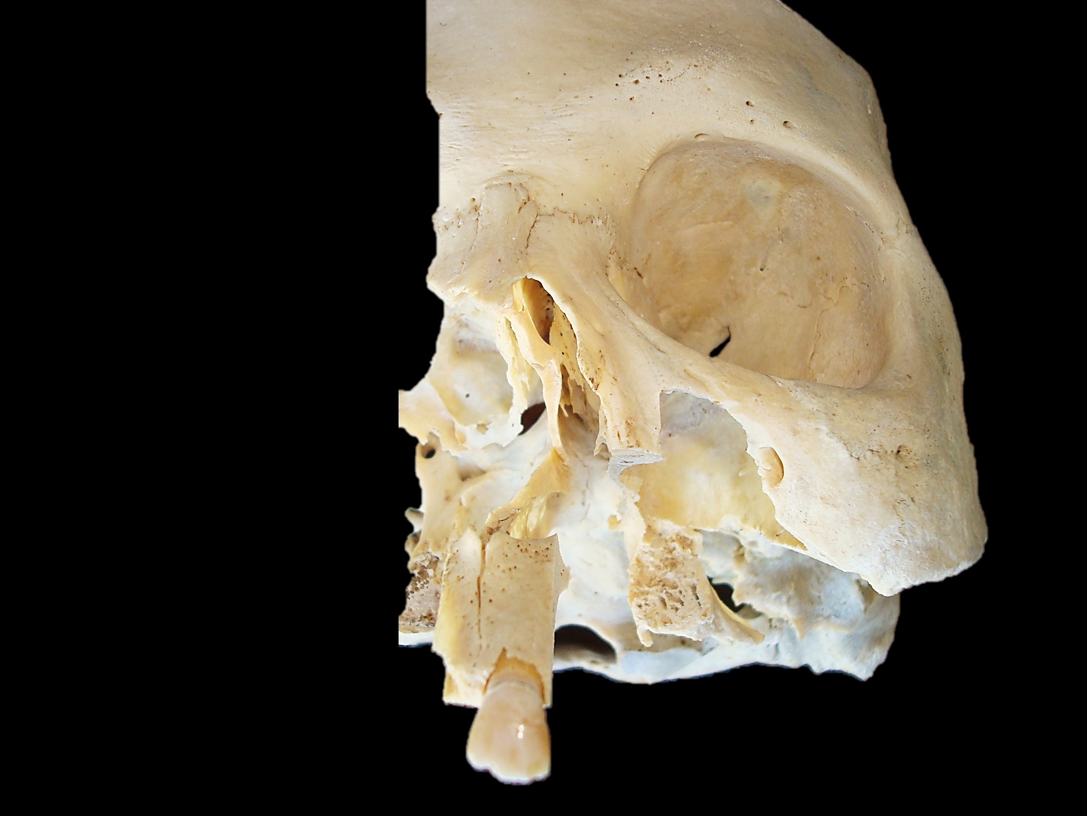
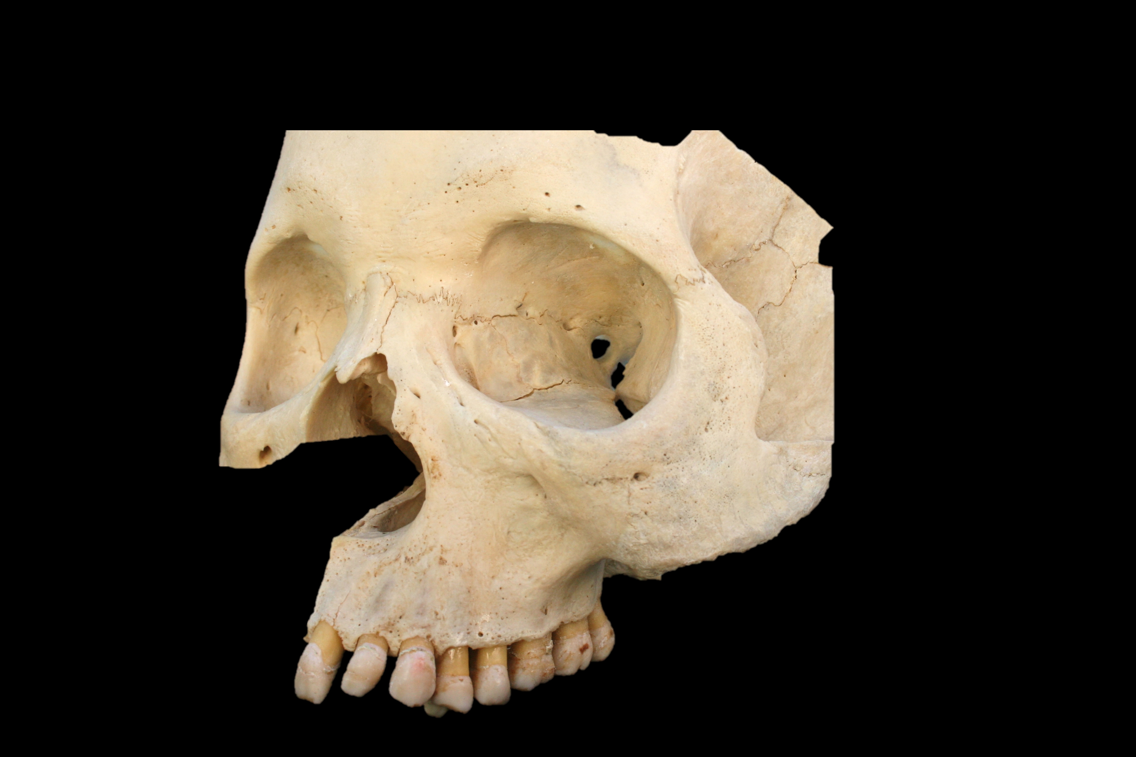
Figures 2, 3 & 4 illustrate the detailed bony anatomy relevant to maxillectomy. Critical surgical landmarks to note include:
The floor of the anterior cranial fossa (fovea ethmoidalis and cribriform plate) corresponds with anterior and posterior ethmoidal foramina located, along the frontoethmoidal suture line
The proximity (5-11mm) of posterior ethmoidal foramen and artery to the optic nerve within the optic foramen
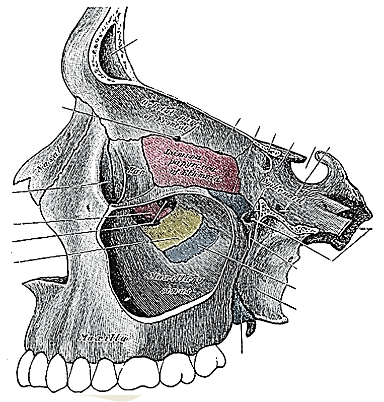
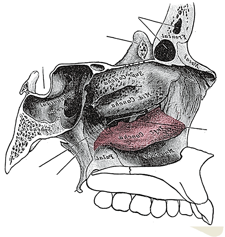
Figure 2 illustrates the bony anatomy of the lateral wall of the nose. The inferior turbinate (concha) may be resected with inferior maxillectomy, but the middle turbinate is preserved.
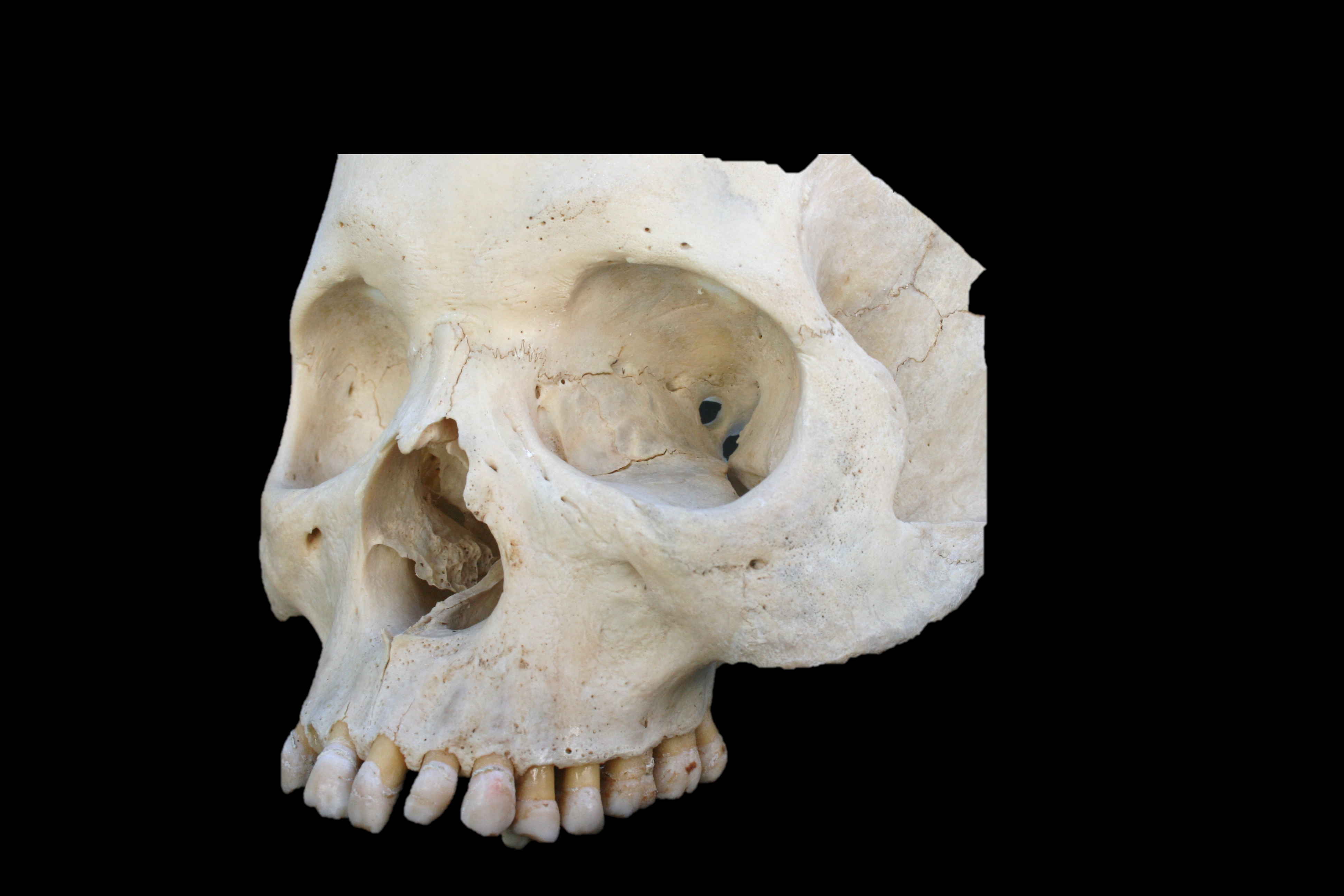
Figure 2: Lateral view of maxilla with windows cut in lateral and medial walls of maxillary sinusFigure 3: Bony anatomy of the lateral wall of the noseFigure 4: Bony anatomy in cadaver
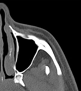
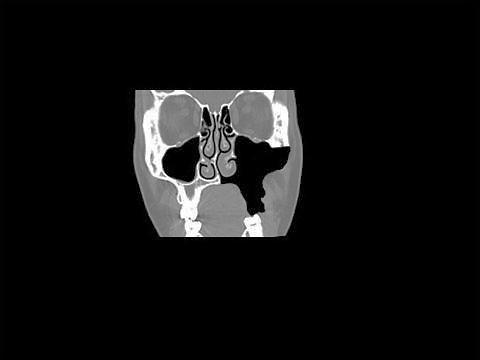
Figure 5 demonstrates the coronal anatomy at the anterior limit of a maxillectomy. Specifically note the lacrimal sac in the lacrimal fossa (Figure 4, 5) which may be transected at surgery, and the relative heights of the floors of the antrum and the nasal cavity.
Figure 5: Coronal CT through lacrimal fossa
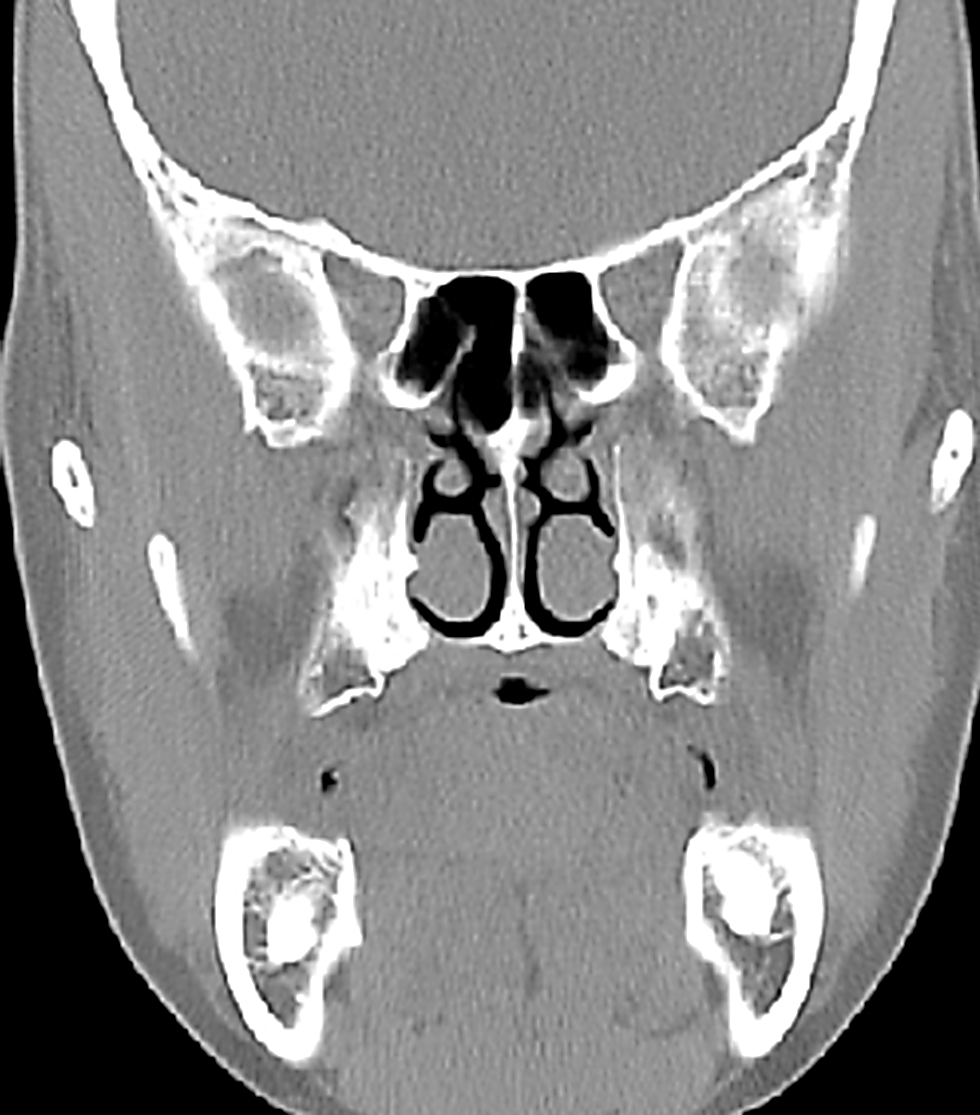
Figure 6 demonstrates the coronal anatomy midway back along a maxillectomy. Specifically note the infraorbital nerve in the orbital floor, the thin lamina papyracea and the relative heights of the floors of the antrum and the nasal cavity.
Figure 6: Anatomy in the coronal plane through the anterior ethmoids midway along a maxillectomy
Figures 7 & 8 demonstrate the value of using the anterior and posterior ethmoidal arteries and frontoethmoidal suture line (Figure 4) to determine the level of the floor of the anterior cranial fossa when opening the lamina papyracea from the orbital side during medial or total maxillectomy.
Figure 7: Note the position of the anterior ethmoidal artery where it passes through its foramen which is located in the frontoethmoidal suture line Figure 8: Coronal slice through posterior ethmoids demonstrating posterior ethmoidal foramen and optic nerve
Figure 9 demonstrates the coronal anatomy immediately posterior to the maxillary sinus, which is in the plane through which an inferior or total maxillectomy is done, and in which the internal maxillary artery and its branches as well as the sphenopalatine ganglion and its branches are encountered within the pterygopalatine fossa. The pterygopalatine fossa communicates laterally with the infratemporal fossa via the pterygomaxillary fissure, and medially with the nasal cavity via the spheno-palatine foramen.
Figure 9: Coronal cut immediately behind the maxillary sinus through the orbital apex, pterygoid plates and pterygopalatine fossa.
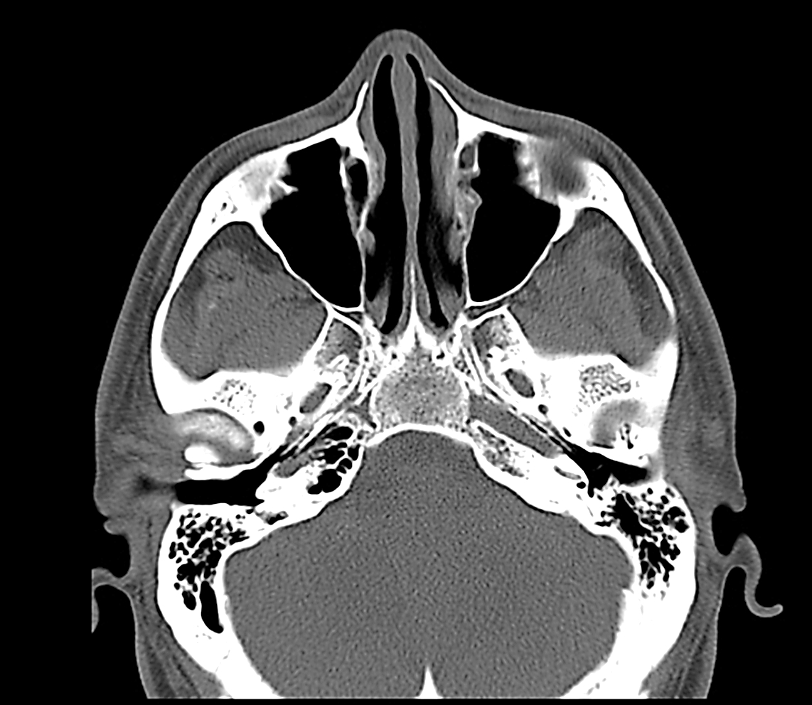
Figures 10 & 11 show axial views of the anatomy of the maxillary sinus. The posterior resection lines of total and inferior maxillectomies pass through the pterygopalatine fossa and pterygomaxillary fissure and the anterior aspect of the pterygoid plates.
Figure 10: Axial cut at level of infraorbital nerve and orbital floorFigure 11: Axial cut at level of infraorbital foramen and pterygoid plates
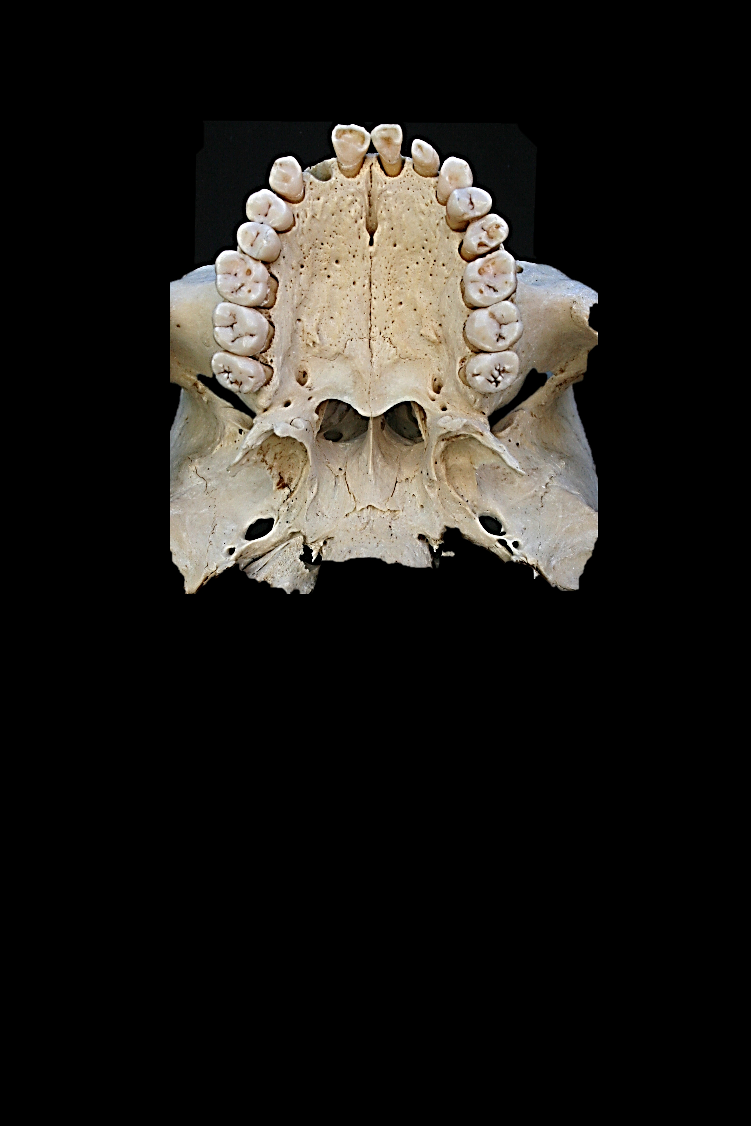
The bony anatomy of the hard palate is illustrated in Figure 12.
Figure 12: Anatomy of hard palate
Vasculature
An understanding of the blood supply of the maxilla permits the surgeon to anticipate when and where to encounter bleeding, and to plan the sequence of the surgery to reserve the bloodier parts of the surgery until last so as to minimise blood loss and to avoid blood obscuring the surgical field.
The only significant vein encountered during maxillectomy is the angular vein(Figure 13) at the medial canthus.
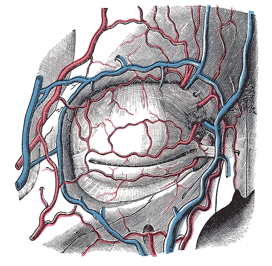
Figure 13: Vasculature around the orbit
The arterial blood supply to the maxilla and paranasal sinuses originates from both the external and internal carotid artery systems. During inferior maxillectomy one can expect to encounter some bleeding from the descending palatine artery, which originates from the maxillary artery in the pterygopalatine fossa, passes inferiorly through the pterygopalatine canal, and emerges from the greater palatine foramen as the greater palatine artery to supply the hard palate.
The arterial supply relevant to inferior maxillectomy is as follows:
Facial/external maxillary artery, a branch of the external carotid artery, courses in the soft tissues of the face and past the medial canthus as the angular artery (Figures 13 & 14)
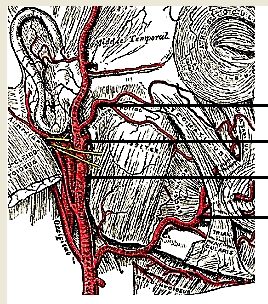
Figure 14: Facial artery and origin of internal maxillary artery, both branches of the external carotid artery
Internal maxillary artery, a branch of the external carotid artery (Figures 14 & 15), passes through the pterygo-maxillary fissure to enter the pterygopalatine fossa.
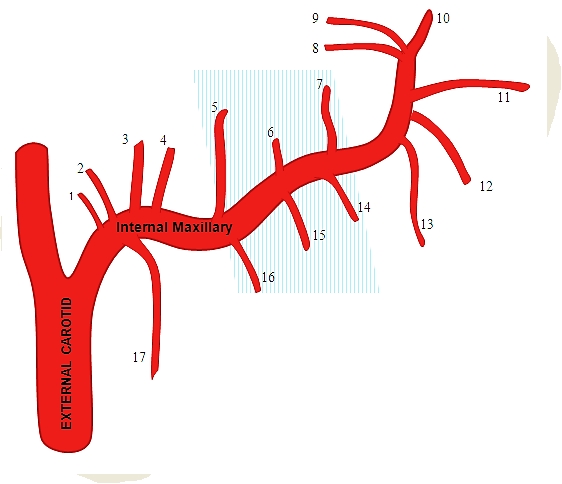
Figure 15: Branches of internal maxillary artery; blue shaded area is the 2nd part of artery before it enters the pterygopalatine fossa
Branches of the internal maxillary arteryof surgical significance include:
Greater palatine artery (descending palatine) (Figure 15): It passes inferiorly from the pterygopalatine fossa through the pterygopalatine canal (Figure 2) and emerges from the greater palatine foramen of the hard palate (Figure 12). It then runs anteriorly medial to the superior alveolus and enters the incisive foramen (Figure 12)
Infraorbital artery: It courses in the floor of the orbit/roof of antrum in the infraorbital groove and canal with the infraorbital nerve and exits anteriorly from the infraorbital foramen to supply the overlying soft tissues of the face (Figures 13 & 15)
Sphenopalatine artery (Figure 15): It enters the nasal cavity through sphenopalatine foramen at the back of the superior meatus
Posterior lateral nasalarteries: Theseoriginate from the sphenopalatine artery after passing through the sphenopalatine foramen
Posterior septal artery: This is a branch of the sphenopalatine artery and crosses the posterior nasal cavity just above the posterior choana to end on the nasal septum; one branch descends in a groove in the vomer to enter the incisive canal and anastomose with the greater palatine artery
Nerves
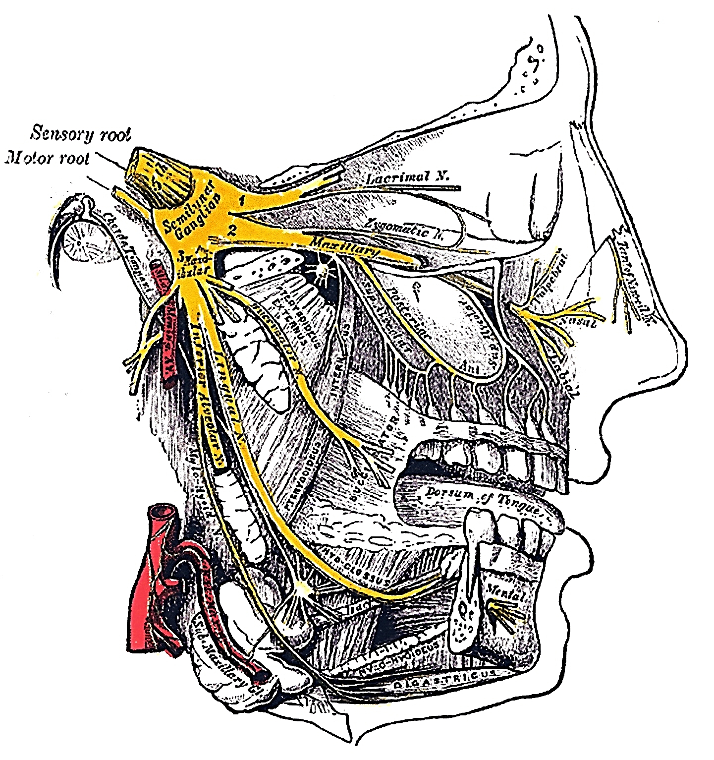
The maxillary division of V (V2) enters the pterygopalatine fossa via the foramen rotundum. The only branch of surgical significance is the infraorbital nerve. It runs in the floor of the orbit/roof of the antrum to exit from the infraorbital foramen (Figure 16).
Figure 16: V2, pterygopalatine ganglion and infraorbital nerve
Inferior Maxillectomy
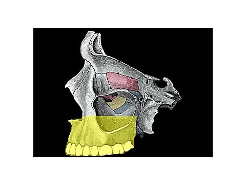
Inferior maxillectomy is employed with tumours limited to the palate and floor of the maxillary sinus and nasal cavity. It entails resection of the hard palate and may include the walls of the maxillary sinus and nasal floor and inferior turbinate, but spares the orbital floor and ethmoid sinuses (Figures 17).
Figure 17: Yellow area indicates extent of bony resection of inferior maxillectomy
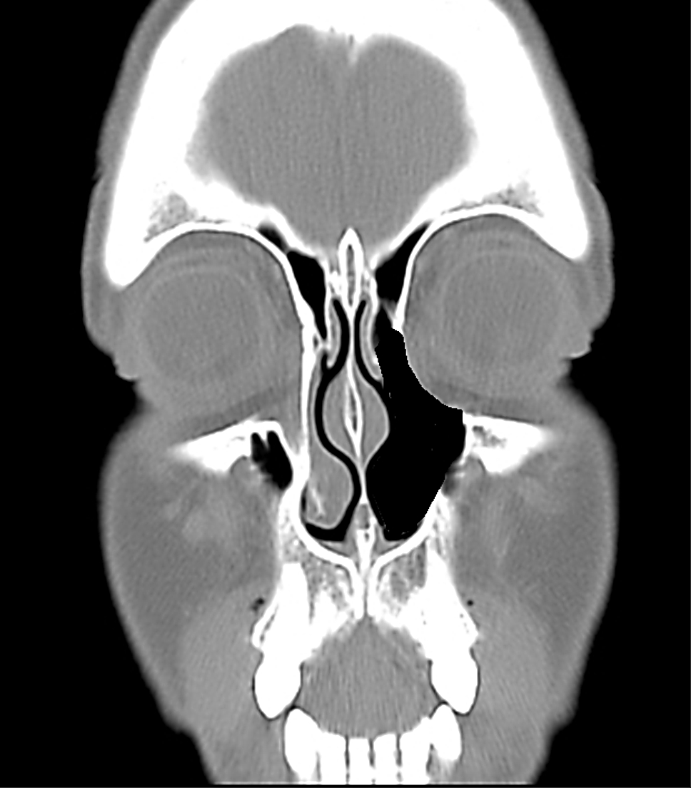
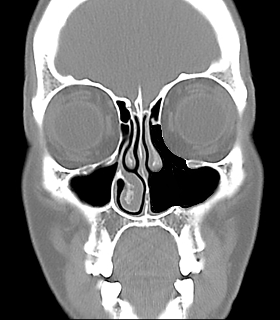
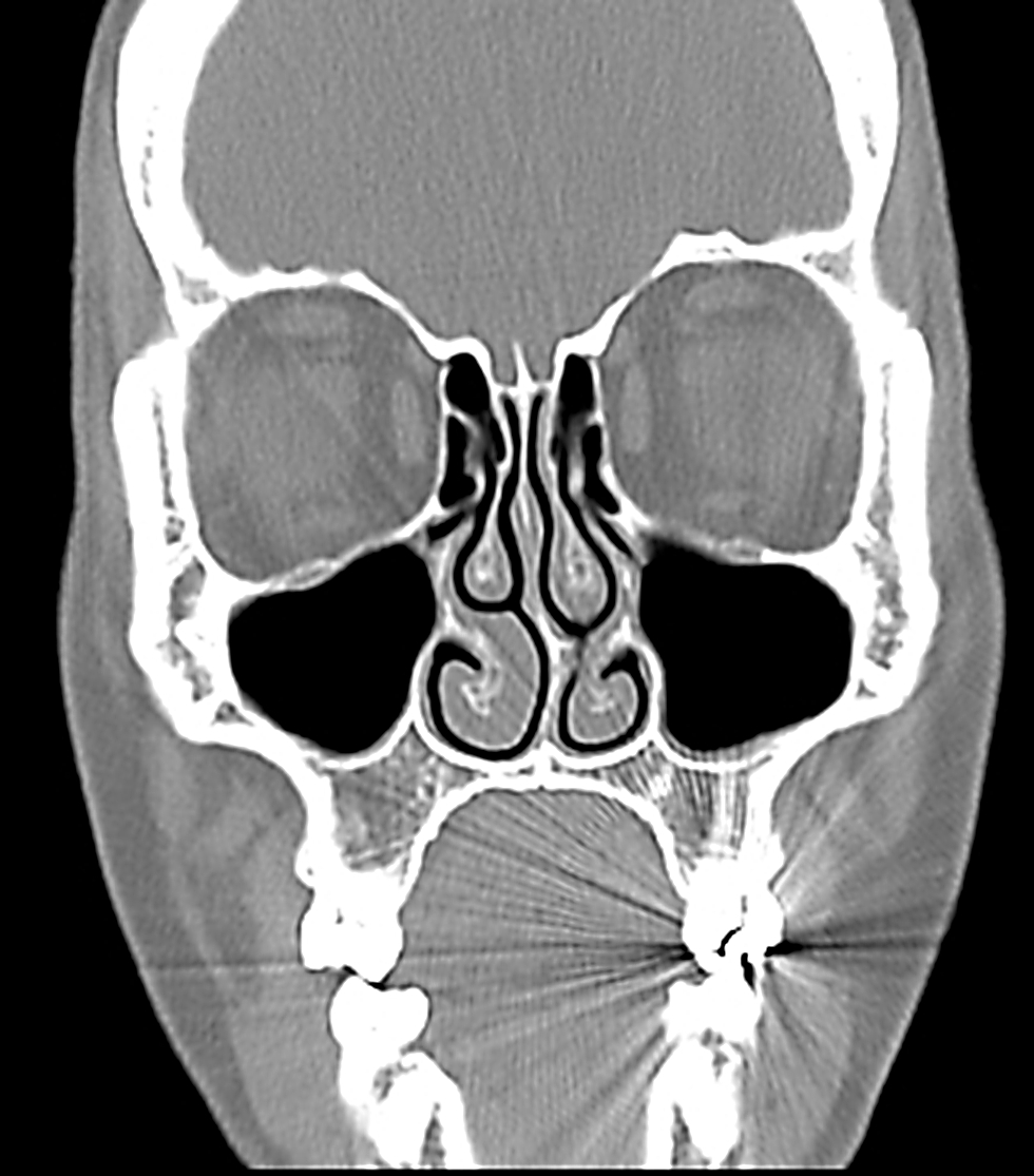
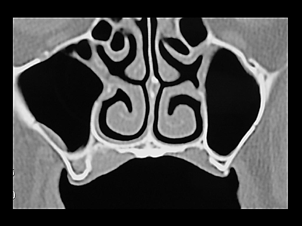
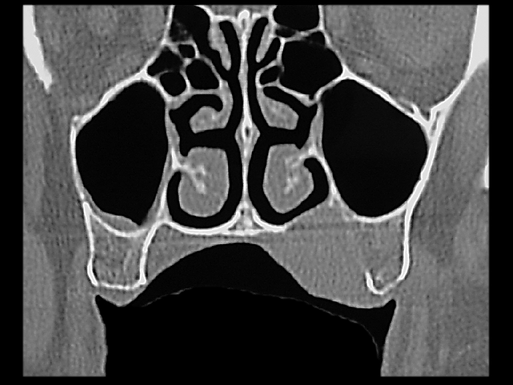
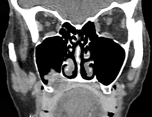
Coronal CT scanning is essential in order to determine the superior extent of a tumour to determine the suitability for an inferior maxillectomy (Figures 18-20).
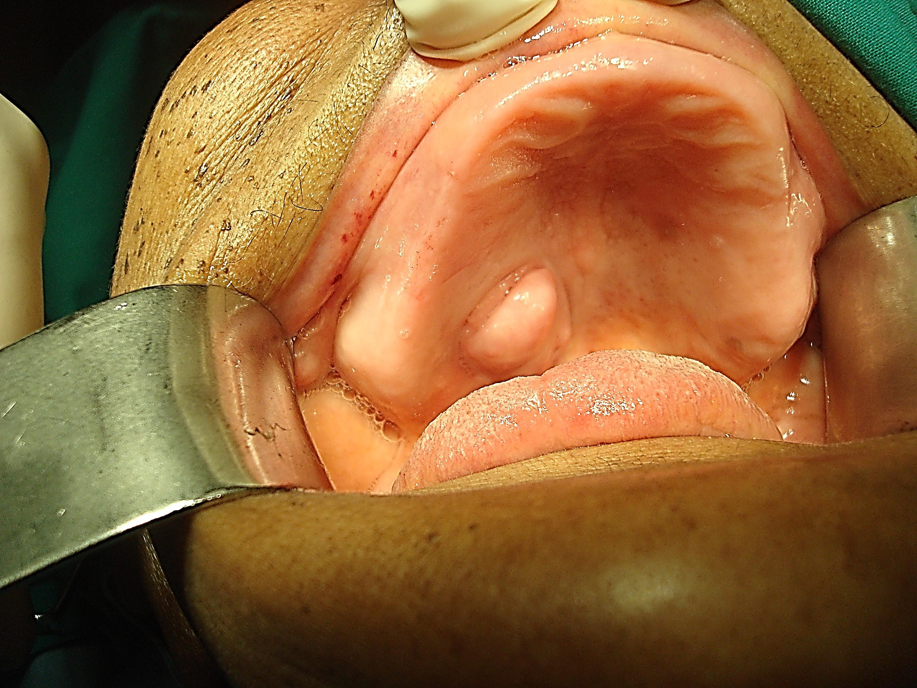
Figure 18: Mucoepidermoid carcinoma of hard palate suited to unilateral inferior maxillectomy with sparing of inferior turbinate and nasal septumFigure 19: Polymorphous low grade adenocarcinoma of palate suited to unilateral inferior maxillectomy with resection crossing midline and including the base of the nasal septumFigure 20: Adenoid cystic carcinoma of hard palate suited to inferior maxillectomy, including inferior turbinate
Surgical steps
The following description refers to a tumour that requires resection of half the hard palate.
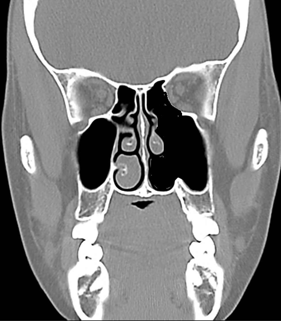
Figure 21 illustrates the extent of the bone resection following a unilateral inferior maxillectomy with preservation of the inferior turbinate.
Figure 21: Coronal CT demonstrating bone removed with unilateral inferior maxillectomy (Inferior turbinate intact)
Preoperative consent includes discussing facial incisions, potential injury to the infraorbital nerve, reconstructive options and the loss of dentition and ability to wear dentures or to have dental implants.
The operation is done under general anaesthesia, with orotracheal intubation, or nasotracheal intubation if only half the palate is to be removed. A temporary tracheostomy is done to ensure an adequate airway in case soft tissue swelling or bleeding occurs. Perioperative broad spectrum antibiotics are administered for 24hrs.
The operation may be considered in 3 stages: soft tissue dissection/bone exposure; bone resection; and closure/reconstruction.Soft tissue dissection/bone exposure
It is important to complete the soft tissue dissection and bone exposure before doing any bone work so as to avoid excessive blood loss.
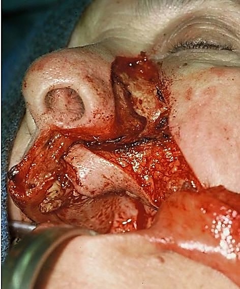
Inferior maxillectomy is done via a sublabial incision or a midfacial degloving approach (Figure 22)
Figure 22: Midfacial degloving approach
Local anaesthetic with vasoconstrictor is injected along the planned mucosal or skin incisions
The sublabial mucosa is incised along the gingivobuccal sulcus with electrocautery
The soft tissues of the face are elevated off the face of the maxilla using cautery or an elevator, remaining hard on bone while doing so. Expose the entire face of the maxilla. Stop the dissection superiorly at the infraorbital foramen taking care to preserve the infraorbital nerve and to avoid troublesome bleeding from the infraorbital artery
Next, free the soft tissues medially from the bone up to the anterior free margin of the nasal aperture with diathermy. Retract the nasal ala and incise the lateral wall of the nasal vestibule to expose the ipsilateral nasal cavity and inferior turbinate, taking care not to injure the inferior turbinate or septum so as to avoid bleeding
Using a tonsil gag in the mouth to retract the tongue, visualise the hard and soft palates and the tumour. Identify the maxillary tuberosity and the bony spines of the pterygoid plates immediately posterior to the tuberosity. Using electrocautery, incise the mucosa of the hard palate along the planned medial resection margin, and extend the sublabial incision laterally around the maxillary tuberosity, and into the groove between the tuberosity and the pterygoid plates.
Palpate and define the posterior edge of the hard palate, and divide the attachment of the soft palate to the hard palate with electrocautery, thereby entering the nasopharynx. Anticipate and coagulate bleeding from branches of the greater and lesser palatine arteries.
At this point the soft tissue dissection is complete
Bony resection (Figs 23-29)
An antrostomy is made in the anterior face of the maxilla with a hammer and gouge or a burr, entering the antrum through the thin bone of the canine fossa (Figure 23). A punch or bone nibbler is used to remove enough bone of the anterior wall of the maxillary sinus to evaluate the tumour extent in the antrum, but taking care to leave a margin of bone around the infraorbital foramen so as to protect the nerve and to avoid bleeding from the infraorbital vessels. Inspect the antrum and determine the extent of the tumour and plan the subsequent bony cuts.
Figure 23: Antrostomy
The inferior maxillectomy can now be done using sharp osteotomes and/or a powered saw. The extent of the bony resection is tailored to the tumour. The sequence of the osteotomies is planned so as to reserve troublesome bleeding to the end. The sequence may have to be adjusted depending on the location and extent of the tumour.
Perform an osteotomy through thelateral wall of the maxillary sinus with an osteotome, bone nibbler or powered saw (Figures 24 -26) up to its junction with the posterior antral wall.
Figure 24: Anterior view of osteotomiesFigure 25: Lateral view of osteotomies, including through pterygomaxillary fissureFigure 26: Osteotomies including osteotomy between maxillary tuberosity and pterygoid plates and of palate
Perform an osteotomy through the anterior medial wall of the maxillary sinus up to the nasal vestibule with an osteotome, bone nibbler or powered saw (Figures 24).
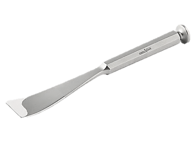
Free the pterygoid plates with a curved osteotome (Figure 27) from the maxillary tuberosity along the posterior vertical line shown in Figures 25 & 26.
Figure 27: Curved osteotome
Perform an osteotomy through the anterior medial wall of the maxillary sinus up to the nasal vestibule with an osteotome, bone nibbler or powered saw (Figures 24).
Free the pterygoid plates with a curved osteotome (Figure 27) from the maxillary tuberosity along the posterior vertical line shown in Figures 25 & 26.
Figure 28: Palatal osteotomies. Note osteotomy passes between palate and pterygoid plates
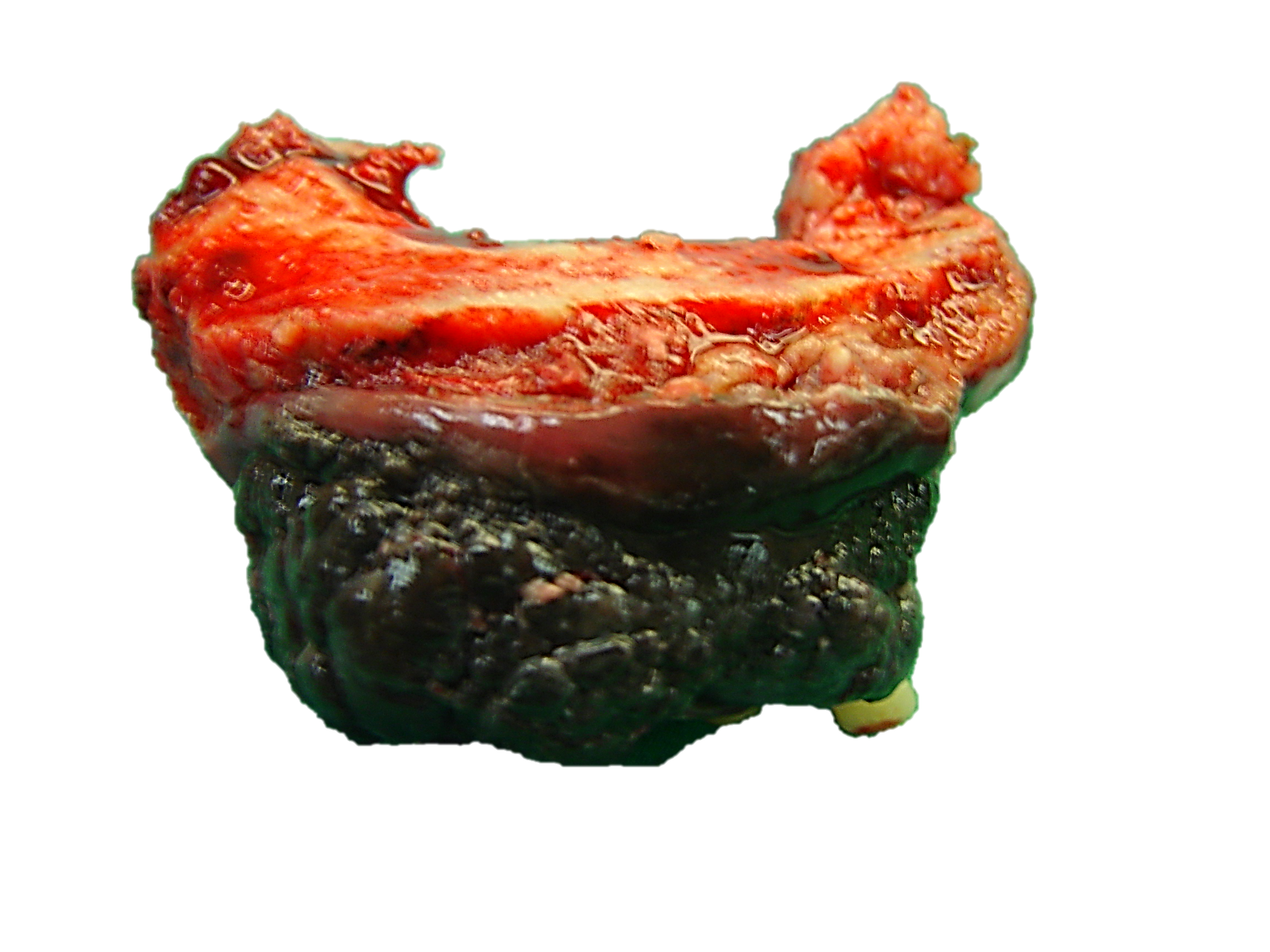
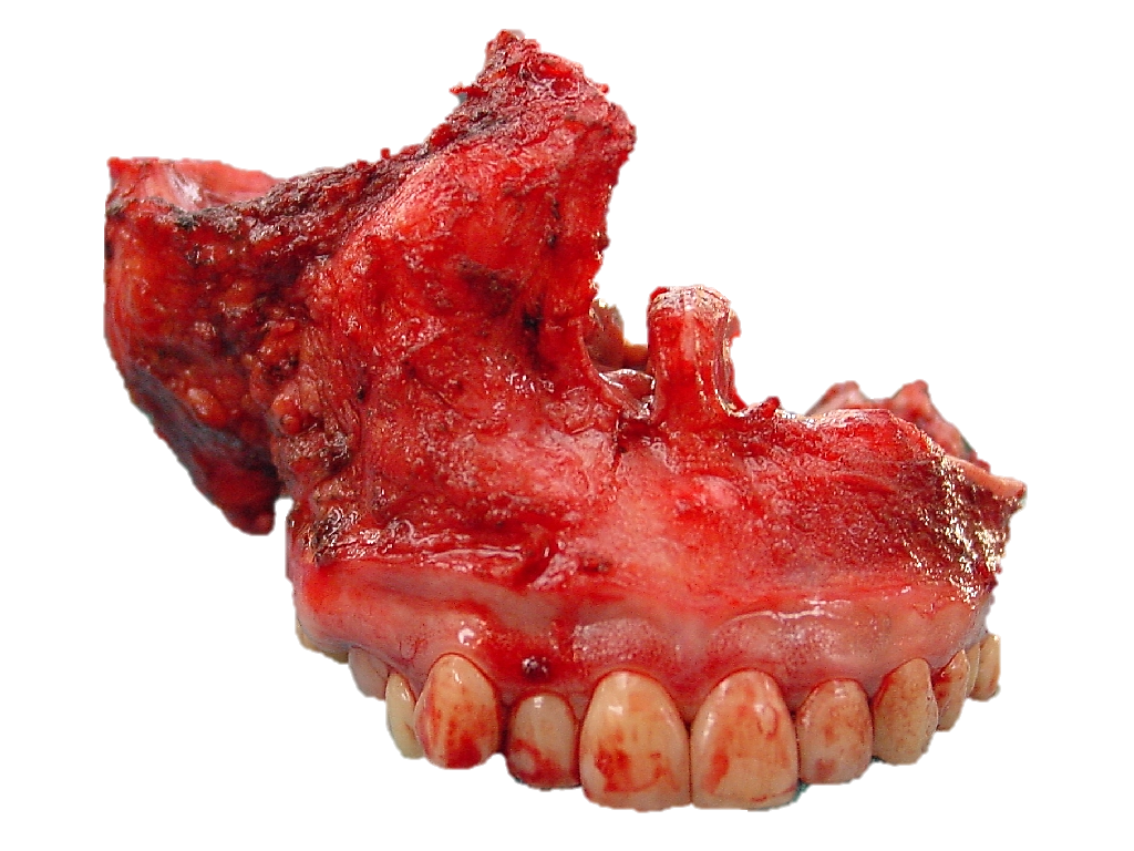
The inferior maxillectomy specimen is then leveraged downwards, fracturing across the posterior antral wall in the process, and the specimen is removed (Figures 29 a, b).
Haemostasis is obtained. The maxillary artery should be looked for as it might have been transected and gone into spasm, and clipped or ligated.
The specimen is inspected to determine the adequacy of the tumour resection margins
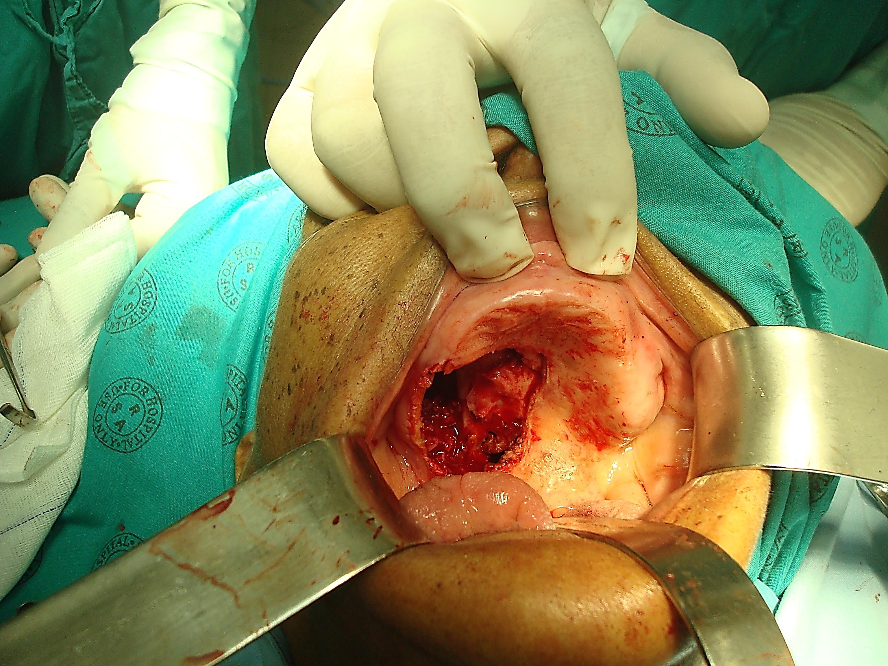
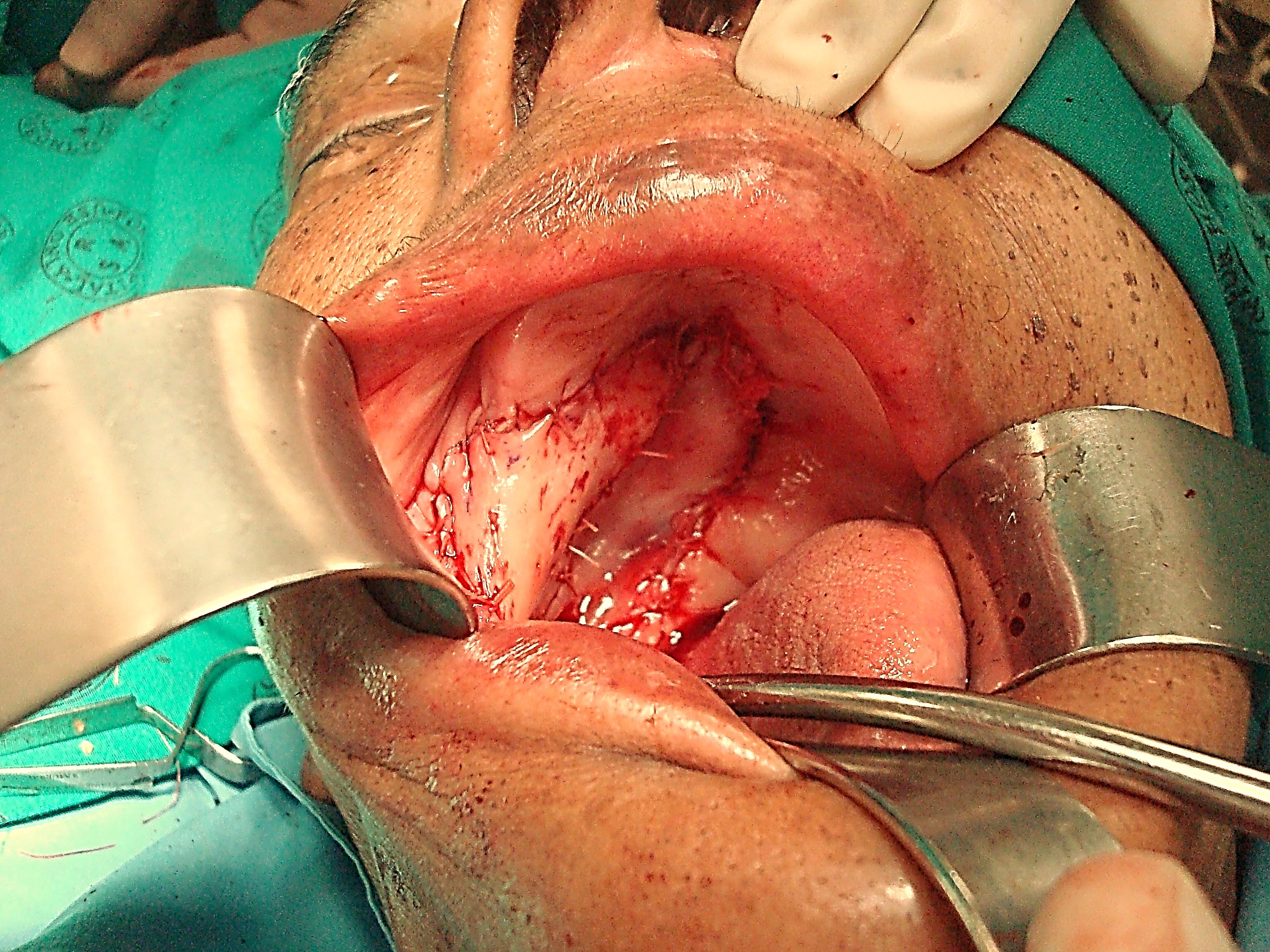
Figures 30 a, b & c show a limited inferior maxillectomy for a minor salivary gland tumour which was reconstructed with a combination of a local rotation flap and a buccinator flap.
Figure 30a: Minor salivary gland tumour of hard palateFigure 30b: Partial inferior maxillectomy suited to an obturator, or reconstruction with buccinator, temporalis muscle, naso-labial or radial free forearm flapsFigure 30c: Partial inferior maxillectomy defect closed with two flaps: buccinator myomucosal and local rotation flaps
With more extensive tumours, bilateral inferior maxillectomy (Figures 1, 31a &, b), or more extensive resections (Figure 32) may be required.
Figure 31a: Malignant melanoma of superior alveolus and hard palateFigure 31b: Bilateral inferior maxillectomy for melanomaFigure 32: (L) inferior maxillectomy with (R) total maxillectomy for sarcoma crossing the midline of the palate
Closure/Reconstruction
The objectives are to restore palatal integrity so as to separate the oral cavity from the nose and antrum, to maintain midfacial projection, and to facilitate dental restoration. This may be achieved in the following ways:
Figure 33: Flap turned in to reconstruct the defectFigure 34: Nasolabial flap inset into the defect and donor site and lip-split closed
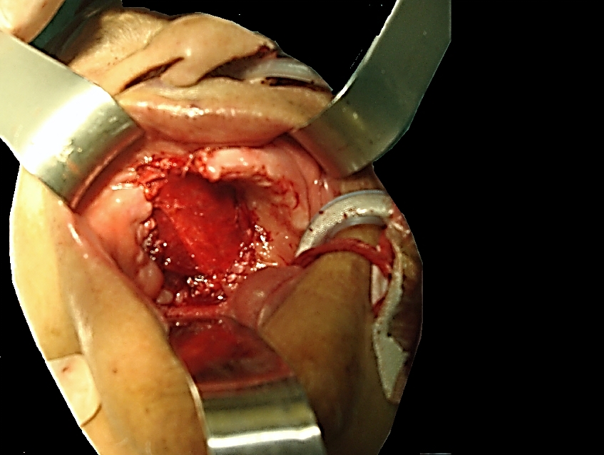
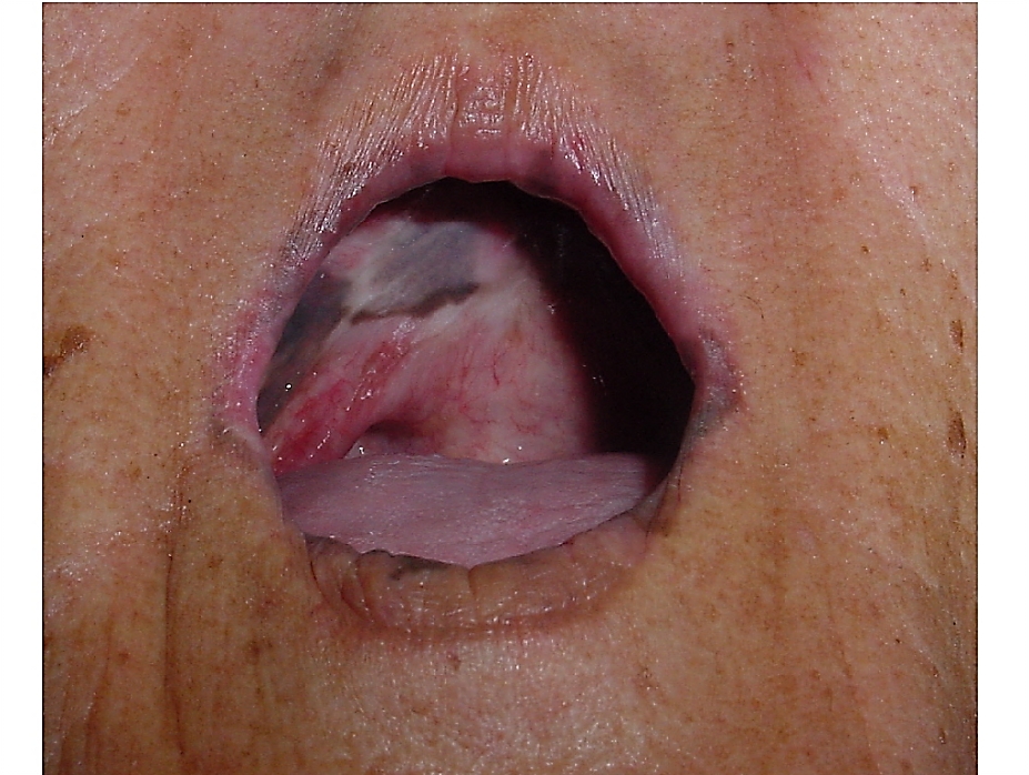
Temporalis muscle flap: This is very well suited, but care has to be taken not to injure the deep temporal arterial pedicle during maxillectomy (Figure 33). A bilateral flap can be used for bilateral inferior maxillectomy defects (Figure 35). It does however preclude the use of dentures
Figure 35: Reconstruction of inferior maxillectomy defect with temporalis muscle flapFigure 36: Mucosalised bilateral temporalis muscle following bilateral inferior maxillectomy
Johan Fagan MBChB, FCORL, MMed
Professor and Chairman
Division of Otolaryngology
University of Cape Town
Cape Town
South Africa johannes.fagan@uct.ac.za