Total maxillectomy refers to surgical resection of the entire maxilla. Resection includes the floor and medial wall of the orbit and the ethmoid sinuses. The surgery may be extended to include orbital exenteration and sphenoidectomy, and resection of the pterygoid plates. It is generally indicated for malignancy involving the maxillary sinus, maxillary bone (sarcomas) and/or orbit and ethmoids, not amenable to lesser or endoscopic resection.
Total maxillectomy is potentially complicated by injury to the orbital contents, lacrimal drainage, optic nerve, ethmoidal arteries, intracranial contents, and may be accompanied by brisk bleeding. A sound understanding of the 3-dimensional anatomy of the maxilla and the surrounding structures is therefore essential. Hence the detailed description of the surgical anatomy that follows.
Surgical Anatomy
Bony anatomy
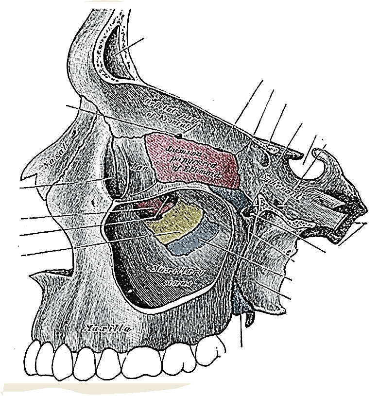
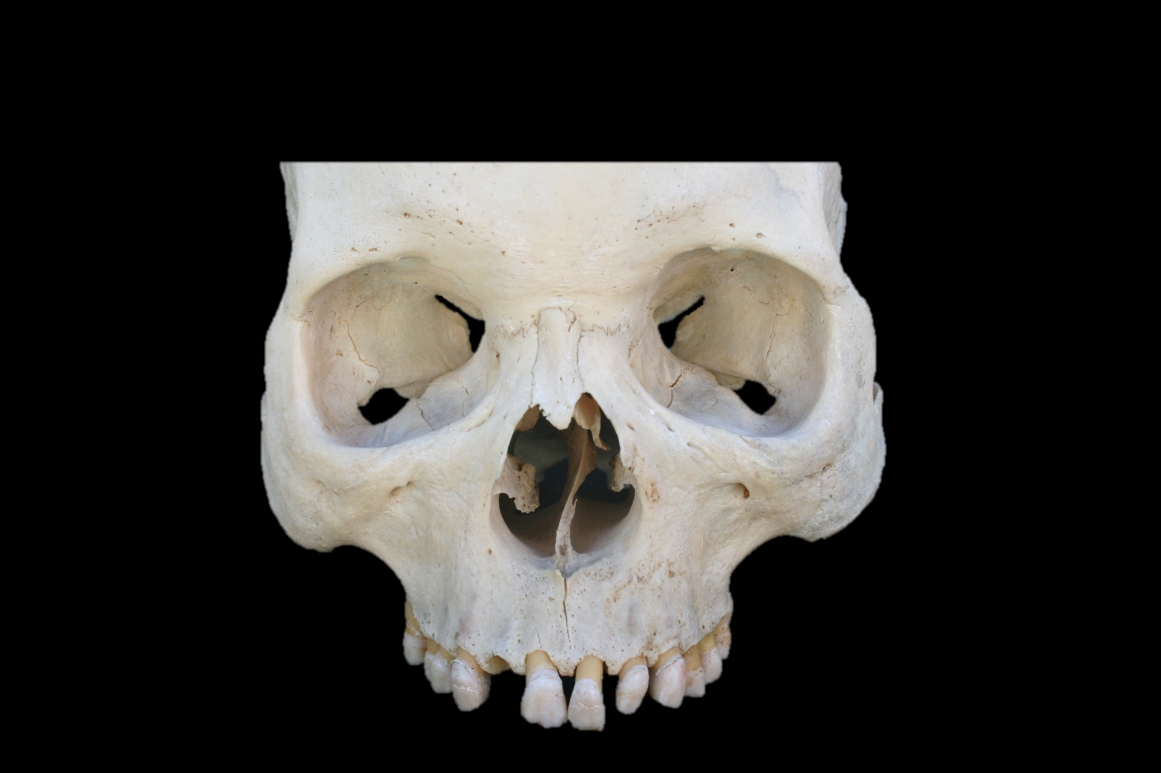
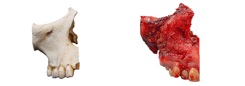
Figures 1 & 2 illustrate the detailed bony anatomy relevant to total maxillectomy. Critical surgical landmarks to note include:
The level of the floor of the anterior cranial fossa (fovea ethmoidalis and cribriform plate) corresponds with anterior and posterior ethmoidal foramina located along the frontoethmoidal suture line
The proximity (5-11mm) of posterior ethmoidal foramen and artery to the optic nerve within the optic foramen
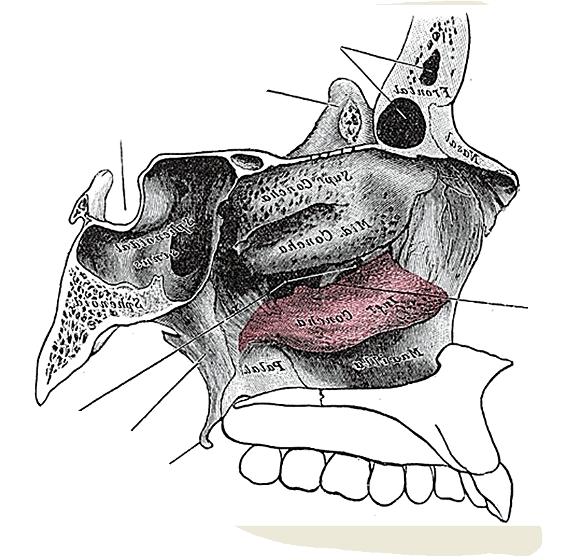
Figure 2 illustrates the bony anatomy of the lateral wall of the nose. The inferior turbinate (concha) is resected with a total maxillectomy, but the middle turbinate is generally preserved, unless involved by pathology.
Figure 1: Lateral view of maxilla with windows cut in lateral and medial walls of maxillary sinusFigure 2: Bony anatomy of the lateral wall of the nose
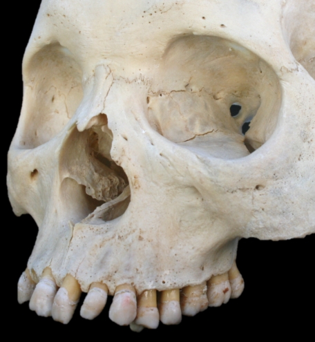
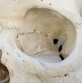
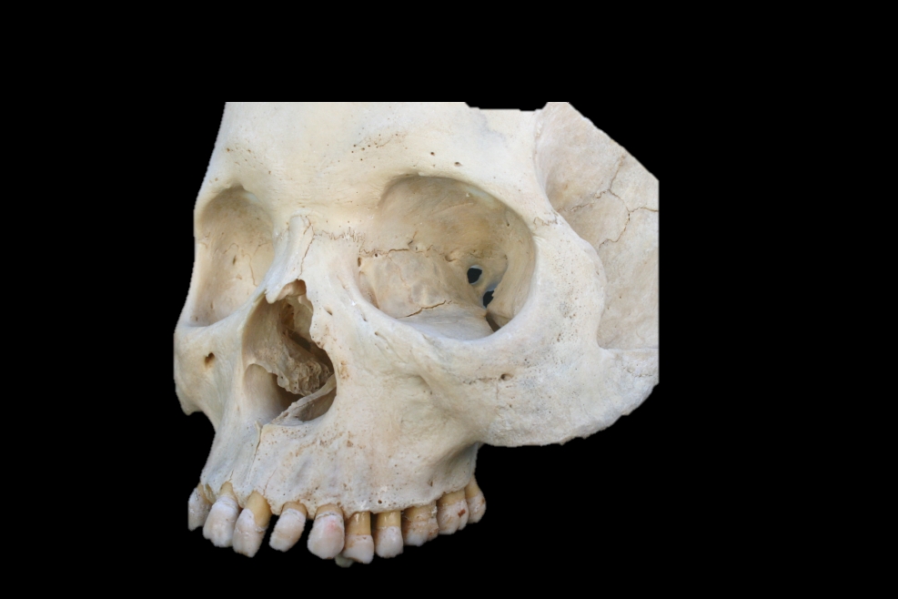
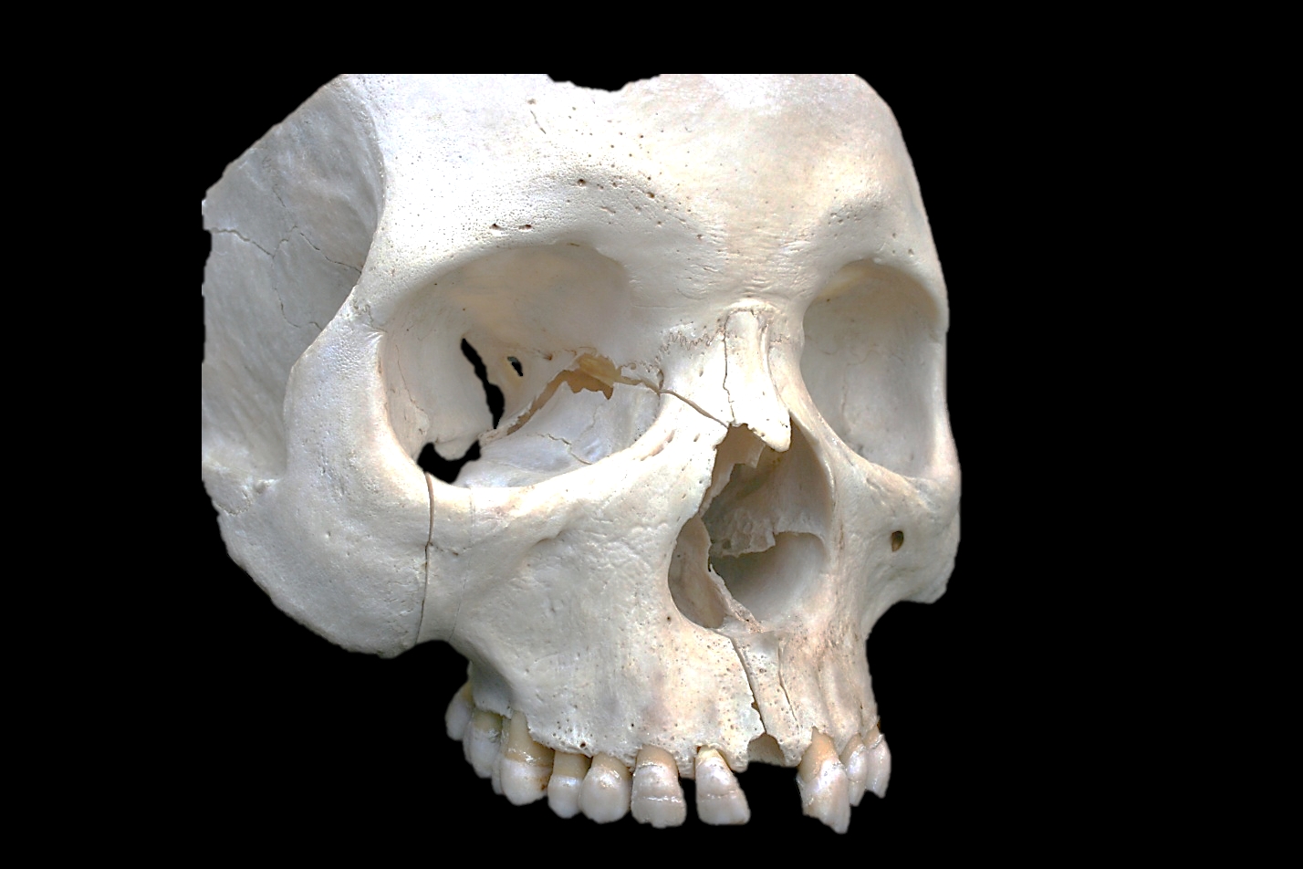
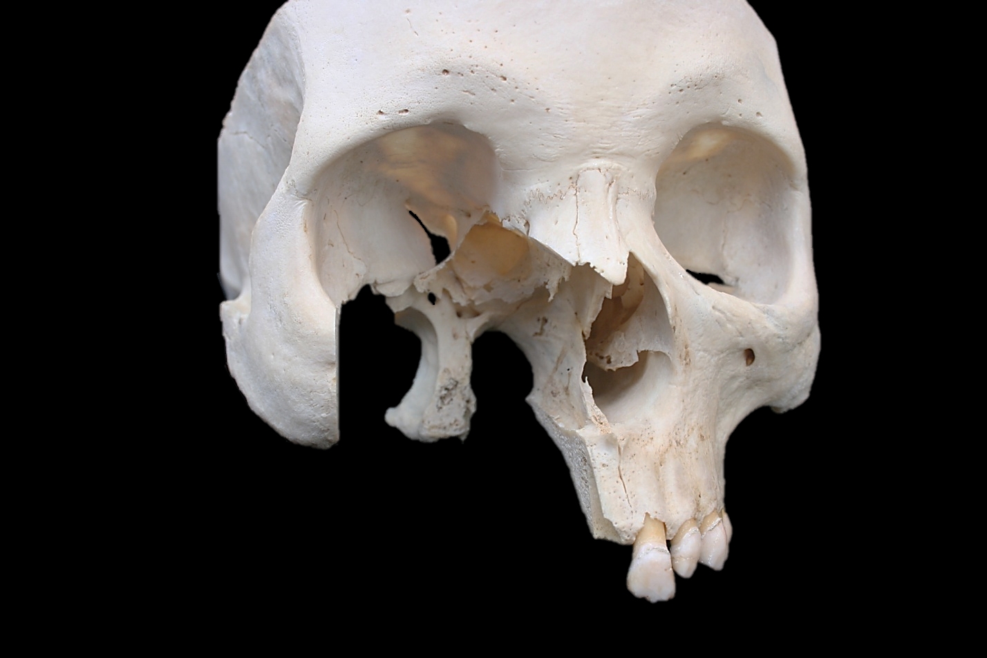
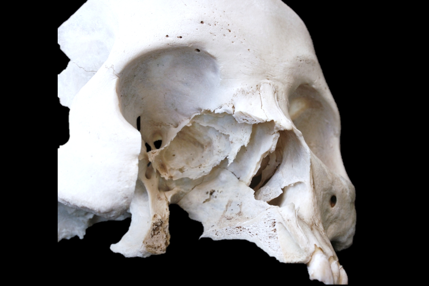
Figure 3 demonstrates the anatomy of the medial wall of the nose in a cadaveric skull. Note in particular the thin lamina papyracea, the lacrimal fossa, the frontoethmoidal suture line and the anterior and posterior ethmoidal foramina and the infraorbital foramen.
Figure 3: Bony anatomy in cadaver
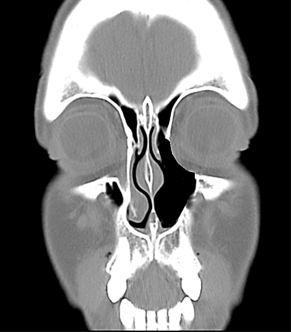
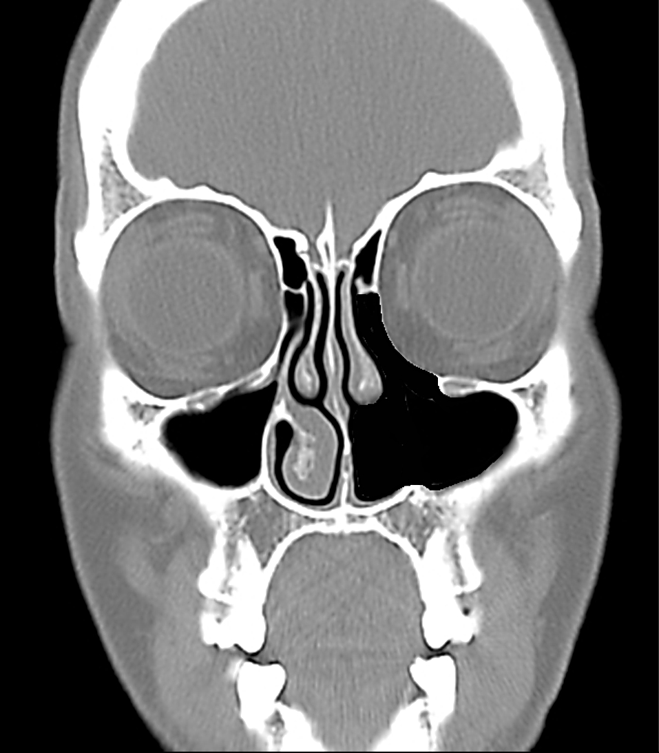
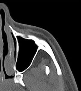
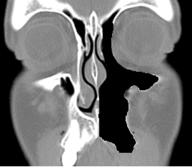
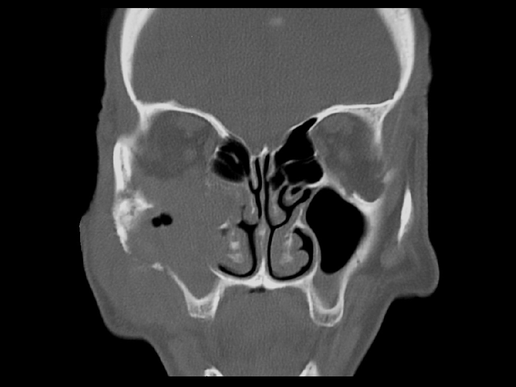
Figure 4 illustrates the coronal anatomy at the level of the anterior extent of a total maxillectomy. Specifically note the lacrimal sac, which is transected at surgery in the lacrimal fossa, and the relative heights of the floors of the antrum and the nasal cavity.
Figure 4: Coronal CT slice through lacrimal fossa
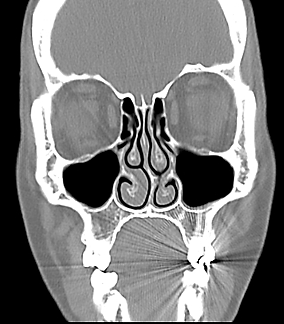
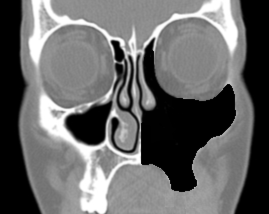
Figure 5 demonstrates the coronal anatomy midway back along a total maxillectomy. Specifically note the infraorbital nerve in the orbital floor, the thin lamina papyracea and the relative heights of the floors of the antrum and the nasal cavity.
Figure 5: Anatomy in the coronal plane through the anterior ethmoids midway along a maxillectomy
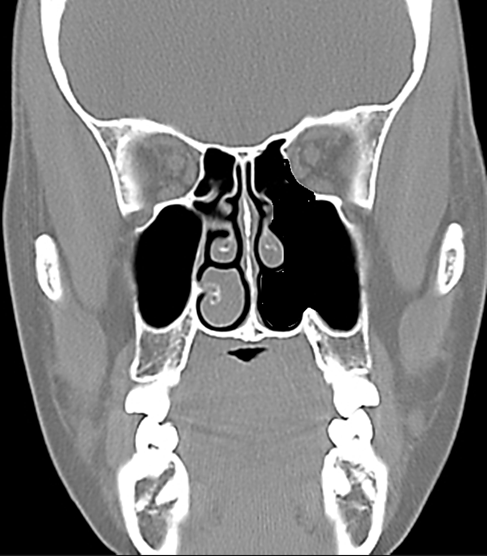
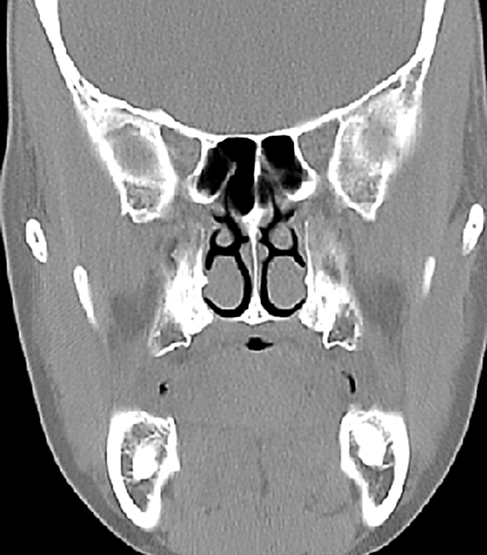
Figures 6 & 7 illustrate the value of using the anterior and posterior ethmoidal arteries and frontoethmoidal suture line to determine the level of the floor of the anterior cranial fossa when opening the lamina papyracea from the orbital side during total maxillectomy.
Figure 6: Note the position of the anterior ethmoidal artery where it passes through its foramen which is located in the frontoethmoidal suture lineFigure 7: Coronal slice through posterior ethmoids demonstrating the posterior ethmoidal foramen and optic nerve
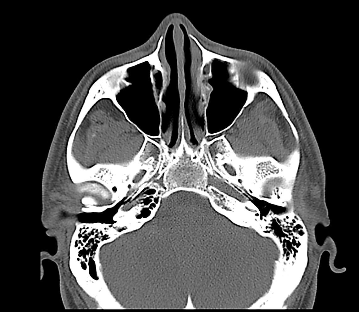
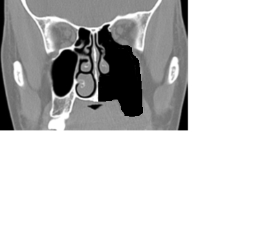
Figure 8 demonstrates the coronal anatomy immediately posterior to the maxillary sinus, which is in the plane through which a total maxillectomy is done, and in which the internal maxillary artery and its branches as well as the sphenopalatine ganglion and its branches are encountered within the pterygopalatine fossa. The pterygopalatine fossa communicates laterally with the infratemporal fossa via the pterygomaxillary fissure, and medially with the nasal cavity via the sphenopalatine foramen.
Figure 8: Coronal cut immediately behind the maxillary sinus through the orbital apex, pterygoid plates and pterygopalatine fossa.
Figures 9 & 10 illustrate axial views of the anatomy of the maxillary sinus. The posterior resection line of total maxillectomy passes through the pterygopalatine fossa and pterygomaxillary fissure and the anterior aspect of the pterygoid plates.
Figure 9: Axial cut at level of infraorbital nerve and orbital floorFigure 10: Axial cut at level of infraorbital foramen and pterygoid plates
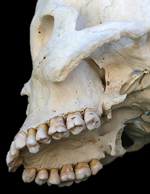
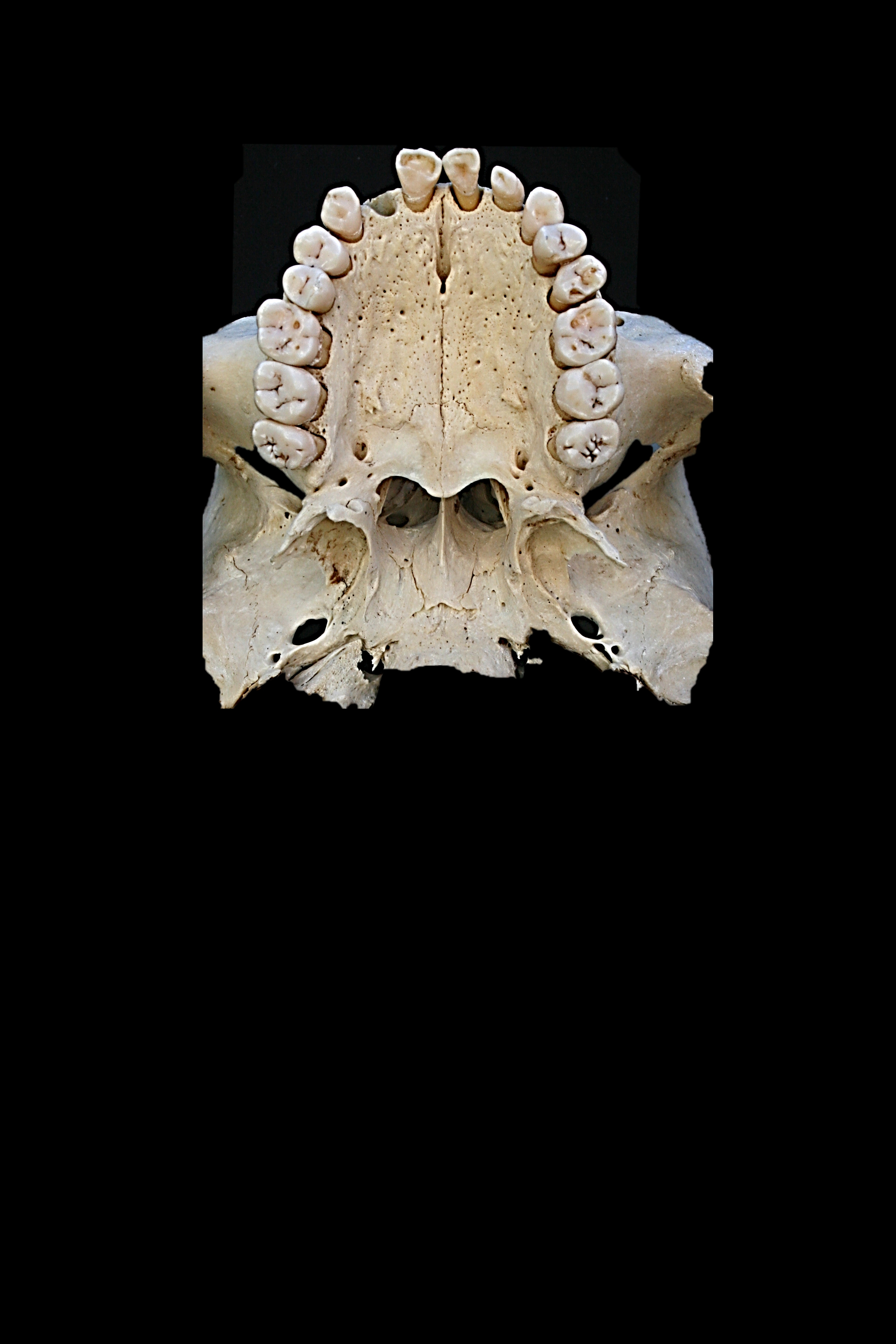
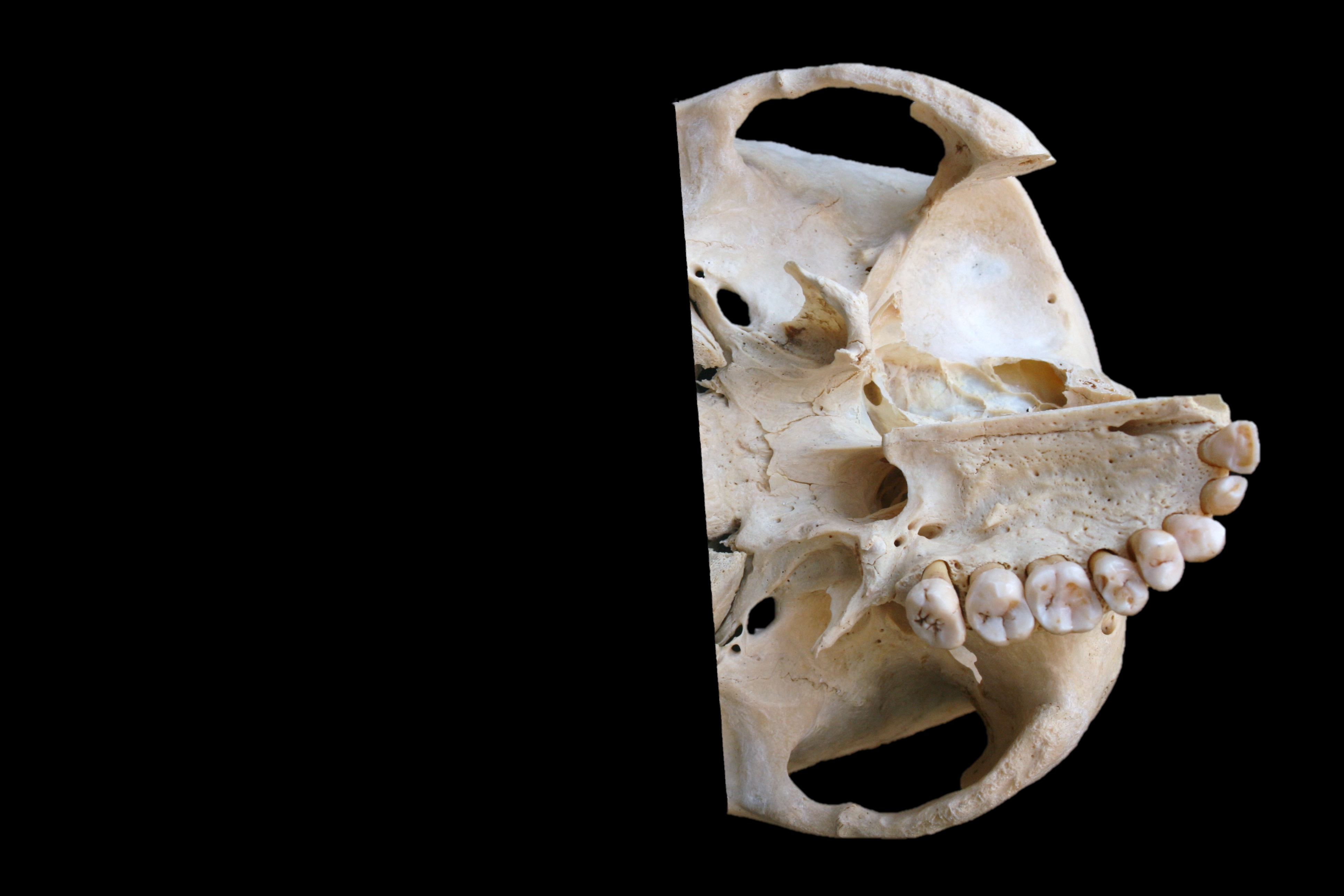
The bony anatomy of the hard palate is illustrated in Figure 11.
Figure 11: Anatomy of hard palate
Vasculature
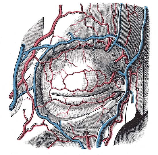
An understanding of the blood supply of the maxilla permits the surgeon to anticipate when and where to encounter bleeding, and to plan the sequence of the surgery to reserve the bloodier parts of the surgery until last so as to minimise blood loss and to avoid blood obscuring the surgical field. The only significant vein encountered during maxillectomy is the angular vein(Figure 12) at the medial canthus.
Figure 12: Vasculature around the orbit
The blood supply to the maxilla and paranasal sinuses originates both from the external (Figures 13, 14) and internal carotid artery systems.
The arterial supply relevant to maxillectomy is as follows:
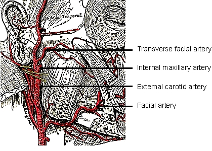
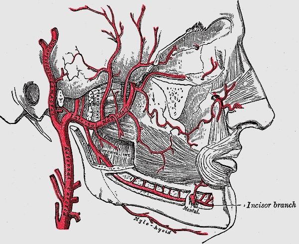
Facial/external maxillary artery, a branch of the external carotid artery (Figures 12, 13)
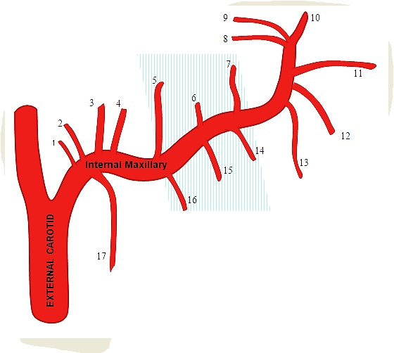
Internal maxillary artery, a branch of the external carotid artery (Figures 13, 14), passes through the pterygo-maxillary fissure to enter the pterygopalatine fossa (Figures 14, 15).
Figure 13: Facial artery and origin of internal maxillary artery, both branches of the external carotid arteryFigure 14: Internal maxillary artery viewed with mandible removed, entering pterygopalatine fossa though the pterygo-maxillary fissureFigure 15: The pterygomaxillary fissure through which internal maxillary artery passes to enter pterygopalatine fossaFigure 16: Branches of internal maxillary artery; blue shaded area is the 2nd part of artery before it enters the pterygopalatine fossa
Branches of the internal maxillary arteryof surgical significance include:
Greater palatine artery (descending palatine) (Figure 16): It passes inferiorly from the pterygopalatine fossa through the pterygopalatine canal (Figure 1) and emerges from the greater palatine foramen of the hard palate (Figure 11). It then runs anteriorly medial to the superior alveolus and enters the incisive foramen (Figure 11).
Infraorbital artery: It courses in the infraorbital groove and canal with the infraorbital nerve in the floor of the orbit/roof of antrum and exits anteriorly via the infraorbital foramen to supply the overlying soft tissues of the face (Figures 12, 14).
Sphenopalatine artery (Figure 16): It enters the nasal cavity through the sphenopalatine foramen at the back of the superior meatus.
Posterior lateral nasalarteries: They are branches of the sphenopalatine artery
Posterior septal artery: This is a branch of the sphenopalatine artery and crosses the posterior nasal cavity just above the posterior choana to end on the nasal septum; one branch descends in a groove in the vomer to enter the incisive canal and anastomose with the greater palatine artery.
Branches of the internal carotid artery of surgical significance are:
Anterior ethmoidal artery: It originates from the ophthalmic artery and enters the orbitthrough the anterior ethmoidal foramen (Figure 3) which is located 25 mm from the anterior lacrimal crest.
Posterior ethmoidal artery: It originates from the ophthalmic artery and enters the orbit through the posterior ethmoidal foramen (Figures 1, 3, 7). It is located approximately 36mm from the anterior lacrimal crest, and 12mm (8-19 mm) from the anterior ethmoidal foramen.
Ophthalmic artery: It emerges with the optic nerve from the optic foramen, 44mm from the anterior lacrimal crest and approximately 6 mm (5-11 mm) from the posterior ethmoidal foramen.
Nerves
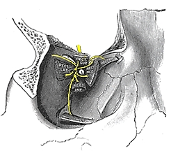
The maxillary division of V (V2) enters the pterygopalatine fossa via foramen rotundum. The only branch of surgical significance is the infraorbital nerve. It runs in the floor of the orbit/roof of the antrum to exit from the infraorbital foramen (Figure 3, 17). The only other major nerve that has to be considered at maxillectomy is the optic nerve.
Figure 17: V2, pterygopalatine ganglion and infraorbital nerve
Orbital structures
Figure 18: Right medial orbital wall
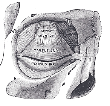
Figure 18 shows the detailed bony anatomy of the orbit. During dissection of the orbit, the following structures are encountered: medial palpebral ligament, orbital septum, lacrimal sac, periosteum, anterior and posterior ethmoidal arteries and inferior orbital fissure (Figure 18, 19). Only when doing orbital exenteration is the superior orbital fissure encountered.
Figure 19: Right orbit showing medial palpebral ligament, orbital septum, lacrimal sac and lacrimal fossa
Orbital septum (Figure 19):This connective tissue structure attaches circumferentially to the periosteum of the orbital margin and acts as a diaphragm that retains the orbital contents. Laterally, it is attached to the orbital margin 1.5mm anterior to the attachment of the lateral palpebral ligament at the lateral orbital tubercle. Superomedially it crosses the supra-orbital groove, passes inferomedially anterior to the trochlea, and follows the posterior lacrimal crest behind the lacrimal sac. It crosses the lacrimal sac to reach the anterior lacrimal crest, passes inferiorly along the anterior lacrimal crest and then laterally along the inferior orbital rim.
Medial palpebral ligament(medial canthal tendon) (Figure 19): This fibrous band fixes the tarsal plates to the medial orbital wall. It is intimately related to the lacrimal drainage system. It lies anterior to the canaliculi, but a deep head inserts into the posterior lacrimal crest and onto the fascia of the lacrimal sac.
Lacrimal sac (Figures 18, 19, 20):It is located in the lacrimal fossa, which is bound medially by the lacrimal bone and the frontal process of the maxilla (Figures 1, 18). It is related anteriorly, laterally, and posteriorly to the medial palpebral ligament.
Figure 20: Right lacrimal system
Inferior orbital fissure (Figure 18, 21): It is situated in the floor of the orbit and separates the sphenoid bone from the maxilla. It transmits the maxillary nerve and a few minor nerves, but no vessels of surgical significance.
Superior orbital fissure (Figure 18, 21): This fissure lies between the lesser and greater wings of the sphenoidbone. Important anatomical structures that pass through the fissure are cranial nerves III, IV, VI; and the superior and inferior divisions of ophthalmic vein.
Figure 21: Superior and inferior orbital fissures of (R) orbit
Total Maxillectomy Operation
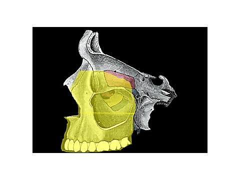
Total maxillectomy entails resection of the entire maxilla, and including the orbital floor and medial wall of the orbit and the ethmoid sinuses (Figure 22). The surgery may be extended to include resection of the lateral orbital wall and zygoma, exenteration of the orbit, sphenoidectomy, and resection of the pterygoid plates.
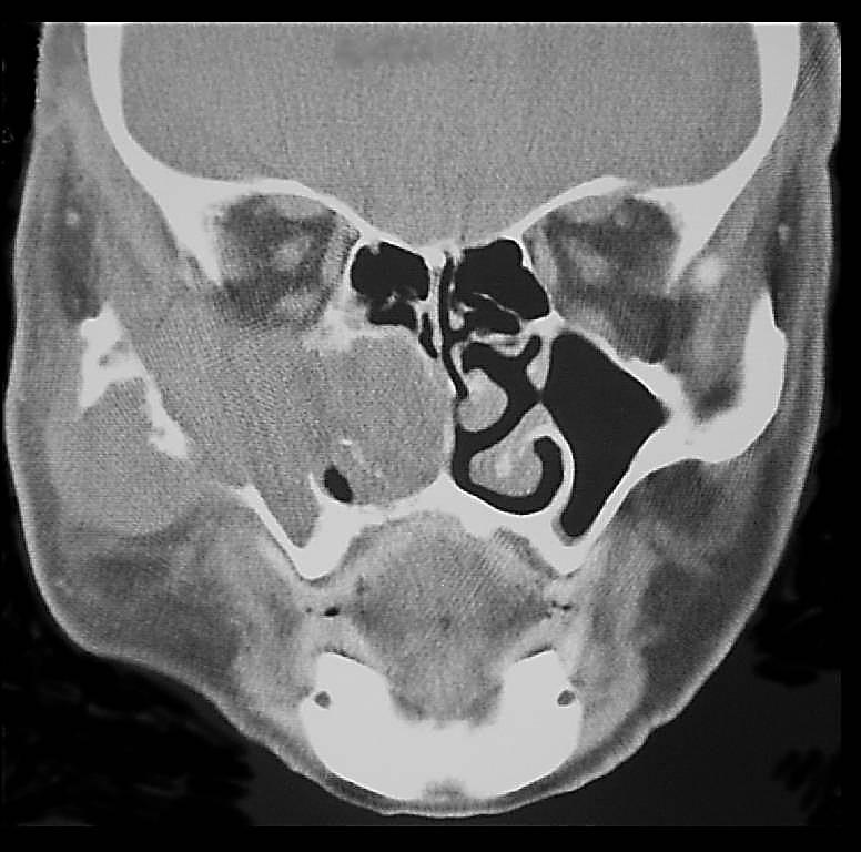
CT scanning is an important means of determining the superior (orbit and roof of ethmoids), posterior (pterygoid plates), lateral (zygoma and infratemporal fossa) and medial extent of the tumour and the resection required.
Figure 22: Yellow area indicates extent of bony resection of total maxillectomy
Once a tumour involves orbital fat and/muscle, then orbital exenteration is generally recommended. Tumour extension to involve the pterygoid muscles precludes a maxillectomy, as getting clear margins once the pterygoid muscles are invaded by cancer is unlikely.
Surgical steps
Preoperative consent includes discussing the need for a tracheostomy, the facial incisions, loss of sensation in the infraorbital nerve distribution, diplopia, epiphora, enophthalmos, telecanthus, potential injury to the optic nerve, and CSF leak.
The operation is done under general anaesthesia, with orotracheal intubation. If the eye is to be preserved then eyelids are sutured together with 6/0 silk taking care not to invert the eyelashes so as to avoid corneal abrasions, unless an orbital exenteration is planned. A tracheostomy is then done. Perioperative broad spectrum antibiotics are administered for 24hrs. Local anaesthetic with vasoconstrictor is injected along the planned skin incisions. The nasal cavity is decongested with a topical vasoconstrictor.
The operation may be considered in 3 stages: soft tissue dissection & bone exposure; bone resection; and closure/reconstruction.
It is important to complete the soft tissue dissection and bone exposure before doing any bone work so as to avoid excessive blood loss.
Soft tissue dissection/bone exposure
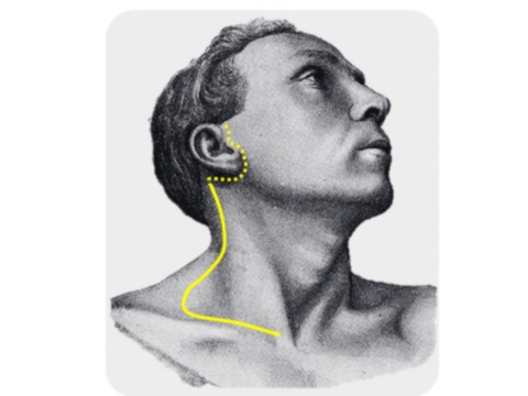
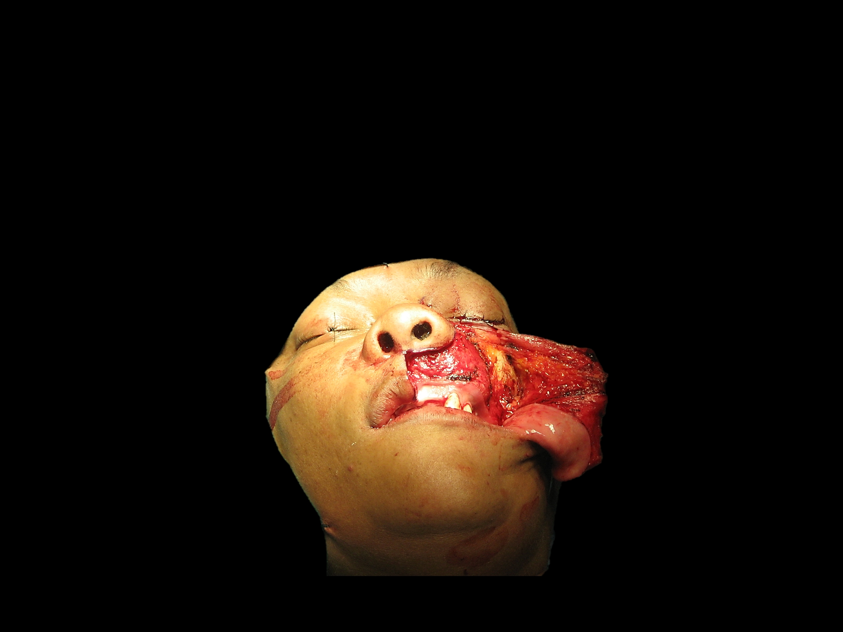
Total maxillectomy may be done via lateral rhinotomy(Figure 23), midfacial degloving (Figure 24)or Weber-Ferguson approach (Figure 25). The midfacial degloving approach avoids facial scars and is suited to resections that do not extend above the orbital floor i.e. do not include resection of the lamina papyracea and ethmoids. Once the resection requires removal of the medial wall of the orbit and the ethmoids, lateral rhinotomy provides better access.
Figure 23: Lateral rhinotomy incision (solid line). A lip split and inferior lid (Weber-Ferguson) incision may be required for accessFigure 24: Midfacial degloving approachFigure 25: Weber-Ferguson approach
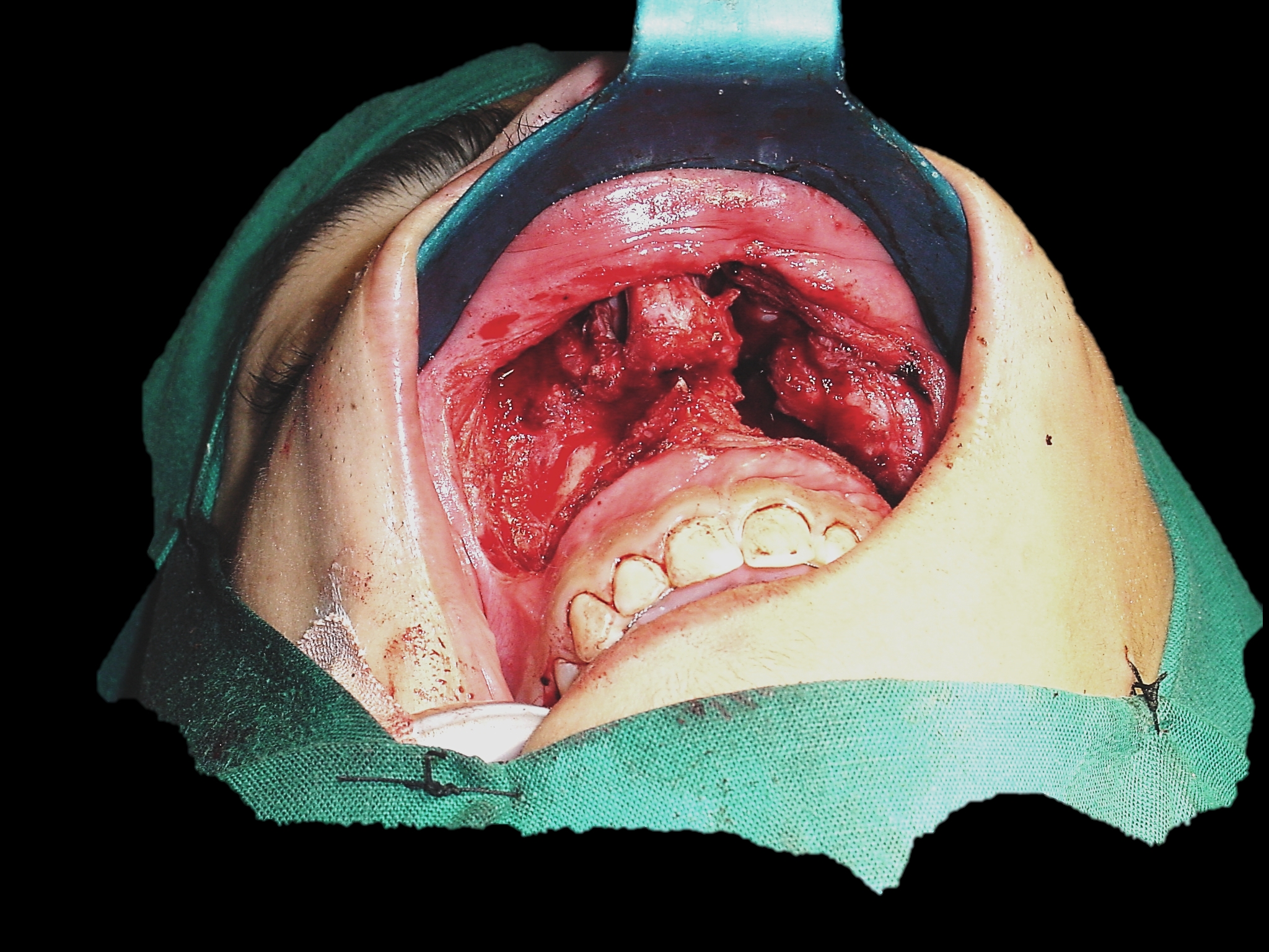
The sublabial mucosa is incised with electrocautery along the gingivobuccal sulcus onto the maxilla and extended all the way to the maxillary tuberosity (Figure 24, 25).
The facial skin is incised with a scalpel. The remainder of the soft tissue dissection may be done with electrocautery. The incision is extended onto the nasal bone and maxilla.
The angular vessels are cauterised or ligated adjacent to the medial canthus of the eye (Figure 12).
With a Weber-Ferguson approach, the lower lid incision is placed close to the palpebral margin so as to avoid oedema of the lower lid above the scar following surgery. The lower lid skin is elevated down to the inferior orbital rim (Figure 25).
The soft tissues of the face are elevated off the face of the maxilla using cautery or an elevator, remaining hard on bone while doing this dissection (Figure 25). Expose the entire face of the maxilla. Transect the infraorbital nerve and vessels with cautery, and if there is concern about perineural invasion, sample the nerve to ensure clear margins. Strip the tissues all the way around the maxilla up to the pterygomaxillary fissure and the zygoma (Figure 15). Do not use sharp dissection beyond the fissure so as to avoid transecting the internal maxillary artery.
Sequentially identify the medial palpebral ligament, anterior lacrimal crest, lacrimal sac in the lacrimal fossa, and posterior lacrimal crest (Figures 18, 19).
Divide the medial palpebral ligament
Elevate the lacrimal sac from its fossa. Transect the sac as distally as possible with a scalpel, so as to facilitate fashioning a dacryocystorhinostomy. Expect some bleeding from the transected sac.
Next the medial and inferior orbit is exposed. Strip the orbital contents in a subperiosteal plane from the lamina papyracea and frontal bone taking care not to fracture or penetrate the paper-thin bone of the lamina papyracea.
Identify the frontoethmoidal suture (Figure 18). This is a crucial surgical landmark as it corresponds with the level of the cribriform plate and the anterior and posterior ethmoidal foramina.
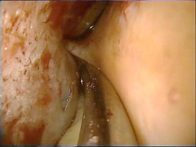
Gently retract the orbital contents laterally and identify the anterior ethmoidal artery as it bridges the divide between the anterior ethmoidal foramen and the periorbita (Figure 26). The anterior ethmoidal artery is ligated, clipped or bipolared and divided (Figure 26, 27), thereby providing access to the posterior ethmoidal artery. It is generally not necessary to divide this vessel.
Figure 26: Weber-Ferguson approachFigure 27: Liga clips being applied to anterior ethmoidal artery (AEA)
Now strip along the floor of the orbit in a subperiosteal plane. Take special care not to tear the periosteum at the inferior orbital margin at the attachment of the orbital septum so as to avoid entering the orbit and causing extrusion of orbital fat.
Next free the soft tissues from the bone up to the anterior free margin of the nasal aperture with diathermy. Retract the nasal ala and incise the lateral wall of the nasal vestibule to expose the ipsilateral nasal cavity and inferior turbinate, taking care not to injure the inferior turbinate or septum so as to avoid troublesome bleeding.
Using a tonsil gag in the mouth to retract the tongue, visualise the hard and soft palates. Identify the maxillary tuberosity and the bony spines of the pterygoid plates immediately posterior to the tuberosity.
Using electrocautery, incise the mucosa of the hard palate along the planned medial resection margin, and extend the sublabial incision laterally around the maxillary tuberosity, and into the groove between the tuberosity and the pterygoid plates.
Palpate and define the posterior edge of the hard palate, and divide the attachment of the soft palate to the hard palate with electrocautery, thereby entering the nasopharynx. Anticipate and diathermise bleeding from branches of the greater and lesser palatine arteries.
At this point the soft tissue dissection is complete. The bony part of the maxillectomy can now been done.
Bony resection
The extent of the bony resection is tailored to the primary tumour and may include the lateral wall of the orbit and zygoma, especially if the antrum is seen to extend into the zygoma on CT imaging. Figures 28 – 30 illustrate the extent of the bone resection following a classic total maxillectomy.
Figure 28: Coronal CT anteriorly though lacrimal fossa, demonstrating resected maxilla, inferior and medial orbital walls and transected lacrimal sacFigure 29: Coronal CT demonstrating resected lateral nasal wall including inferior turbinate and uncinate process, orbital floor including the infraorbital nerve, the lamina papyracea and anterior ethmoidectomy, with preservation of the middle turbinateFigure 30: Coronal CT more posteriorly demonstrating resected maxilla, inferior turbinate, medial and inferior orbital walls, and posterior ethmoidectomy with resection remaining below the level of the posterior ethmoidal foramen, and with preservation of middle
The sequence of the osteotomies is planned to reserve troublesome bleeding to the end of the procedure (Figure 31 - 35). It may need to be adjusted depending on the location and extent of the tumour.
Osteotomy through inferior orbital rim and along orbital floor (Figures 29-31): A sharp osteotome/power saw/bone nibbler is used to cut through the malar buttress / inferior orbital rim. This osteotomy is placed lateral to the antrum as seen on the CT scan so as not to enter the antrum. While retracting and protecting the orbital contents with a narrow copper retractor the osteotomy is then continued posteriorly through the thin bone of the orbital floor/antral roof using a sharp osteotome and aiming for the infraorbital fissure.
Osteotomy through frontal process of maxilla and lacrimal bone (Figures 31-33): This thick bone is best transected with a Kerrison’s rongeur or oscillating saw. There is often persistent minor bleeding from the bone that may be controlled with bone wax or cautery. The osteotomy is directed towards, but kept a few millimetres below, the level of frontoethmoidal suture line.
Osteotomy through lamina papyracea and anterior ethmoids (Figures 6, 18, 28-33): This osteotomy is done by gently tapping on an osteotome to enter the ethmoid air cell system while carefully retracting the orbital contents laterally. It is critical that this osteotomy be kept a few millimetres below the level of the frontoethmoidal suture line and the ethmoidal foramina so as to avoid fracturing or penetrating through the cribriform plate (Figures 5, 6, 29, 39). The osteotomy stops short of the posterior ethmoidal artery and then is directed inferiorly towards the orbital floor so as to safeguard the optic nerve.
Palatal osteotomy (Figures 32-35, 39, 40): A sharp osteotome/power saw is used to cut vertically through the superior alveolus and hard palate. The placement of this osteotomy is dependent on the palatal extent of the tumour. It is often preferable to extract a tooth and to place the osteotomy through the dental socket, rather than to place it between two teeth as this might devitalise the adjacent two teeth and it makes soft tissue closure more difficult. The palatal osteotomy is extended to the posterior margin of the hard palate.
Figure 34: Palatal osteotomies. Note osteotomy passes between palate and pterygoid plates
Osteotomy of nasal septum: This is only required when the palatal osteotomy is placed across the midline. The nasal septum is then divided parallel to the nasal floor with an osteotome or heavy scissors.
Osteotomy to separate maxillary tuberosity from pterygoid plates (Figures 34-36): This is the final osteotomy, and is done by tapping with a (curved) osteotome in the groove between the maxillary tuberosity and the pterygoid bone. Superiorly this cut ends in the pterygomaxillary fissure and the pterygopalatine fossa.
The maxillectomy specimen can now be gently down-fractured.
The internal maxillary artery tethers the specimen laterally, and is clipped and divided where it enters the pterygomaxillary fissure. If the artery is inadvertently transected, it is clipped and ligated. If the artery is not apparent, then it should be specifically looked for as it may have gone into spasm and may bleed later.
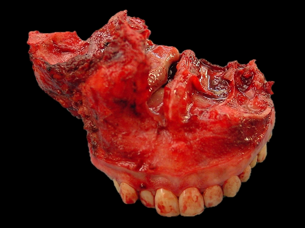
The specimen is removed and inspected to determine the adequacy of the tumour resection (Figure 37).
Figure 37: Maxillectomy specimens
The remnants of the ethmoids are carefully inspected. An external ethmoidectomy may safely be completed up to the cribriform plate (Figures 29, 30, 38-40). The need for external frontoethmoid-ectomy +/- sphenoidectomy is determined, and evidence of a CSF leak is excluded.
Arterial and venous bleeding occur from the pterygoid venous plexus it may initially be controlled with packing. Meticulous haemostasis is achieved with bipolar cautery, suture ligatures, clips, bone wax, and topical haemostatics.
Figures 38-41 demonstrate total maxillectomy defects. Note the location of the face of the sphenoid sinus that may be opened and stripped of mucosa for oncological reasons.
The objectives of closure and reconstruction are to prevent epiphora, to separate the oral cavity from the nose and orbit, to preserve the facial contours, to minimise enophthalmos and diplopia, to maintain a nasal airway, and to restore dentition.
Epiphora: The lacrimal sac is slit along its length and marsupialised by suturing its edges to the surrounding soft tissue with vicryl sutures.
Oronasal separation may be achieved in a number of ways:
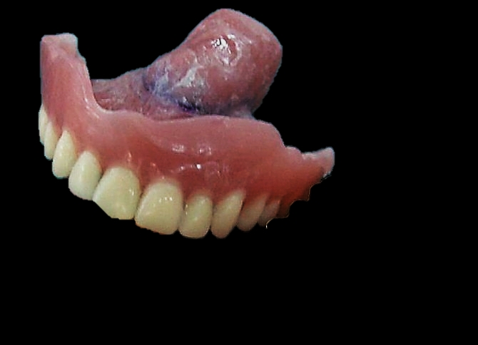
Dental obturator (Figure 42): This can be fashioned to fill the defect and to restore dentition.
Figure 42: Example of obturator
The inner aspect of the maxillectomy cavity is lined with a split-thickness skin graft, which is kept in place with an antiseptic-impregnated gauze pack. The pack is retained with overlying sutures or by screwing a prefashioned plate to the remaining palate with lag screws. A circumferential scar band develops at the interface between the skin graft and the remaining gingivobuccal mucosa and aids to secure the obturator. A temporary obturator may be fitted after the gauze is removed on Day 5. The obturator is remoulded as the cavity heals and contracts.
Temporalis muscle flap: Special care has to be exercised not to injure the deep temporal artery supply to the flap.
Anterolateral free thigh flap: This provides good bulk and palatal skin cover, but precludes wearing a denture.
Scapula free flap: Although it provides oronasal separation the bone is suboptimal for dental implants.
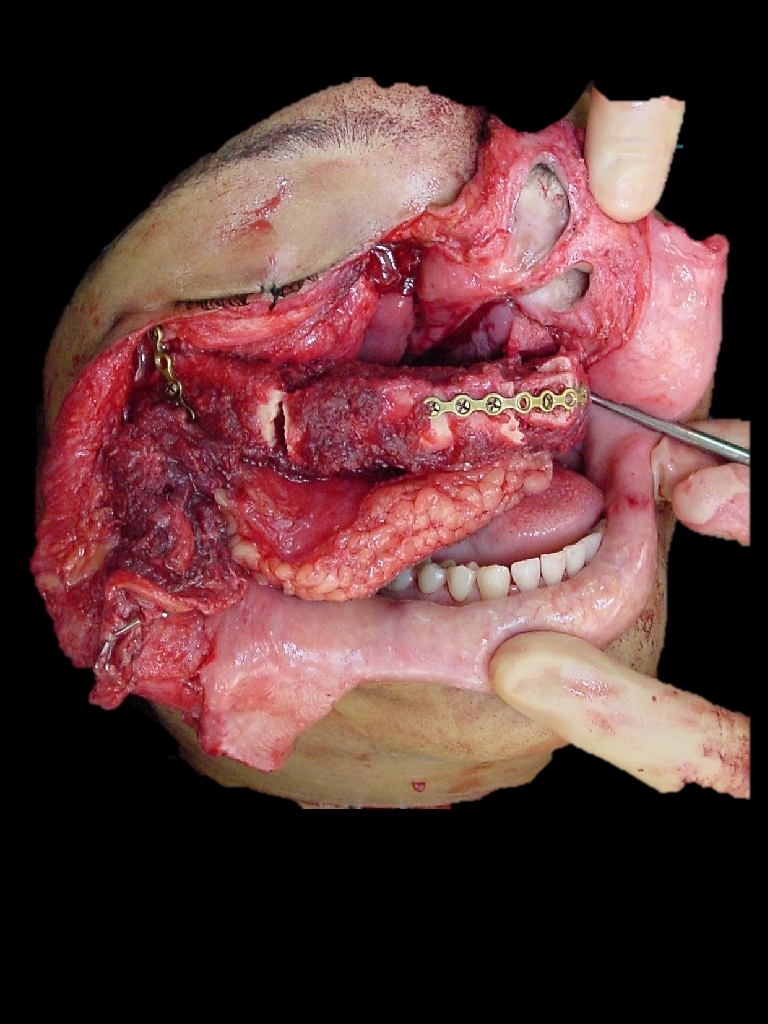
Free fibula flap (Figure 43, 44): This is an excellent option as it permits dental implants.
Dentition: This may be achieved with an obturator (Figure 42) or with dental implants inserted into a free fibula flap.
Figure 43: Free fibula flap reconstruction
Facial contour: Other than the temporalis muscle flap, all the techniques listed above under “oronasal separation” achieve this objective.
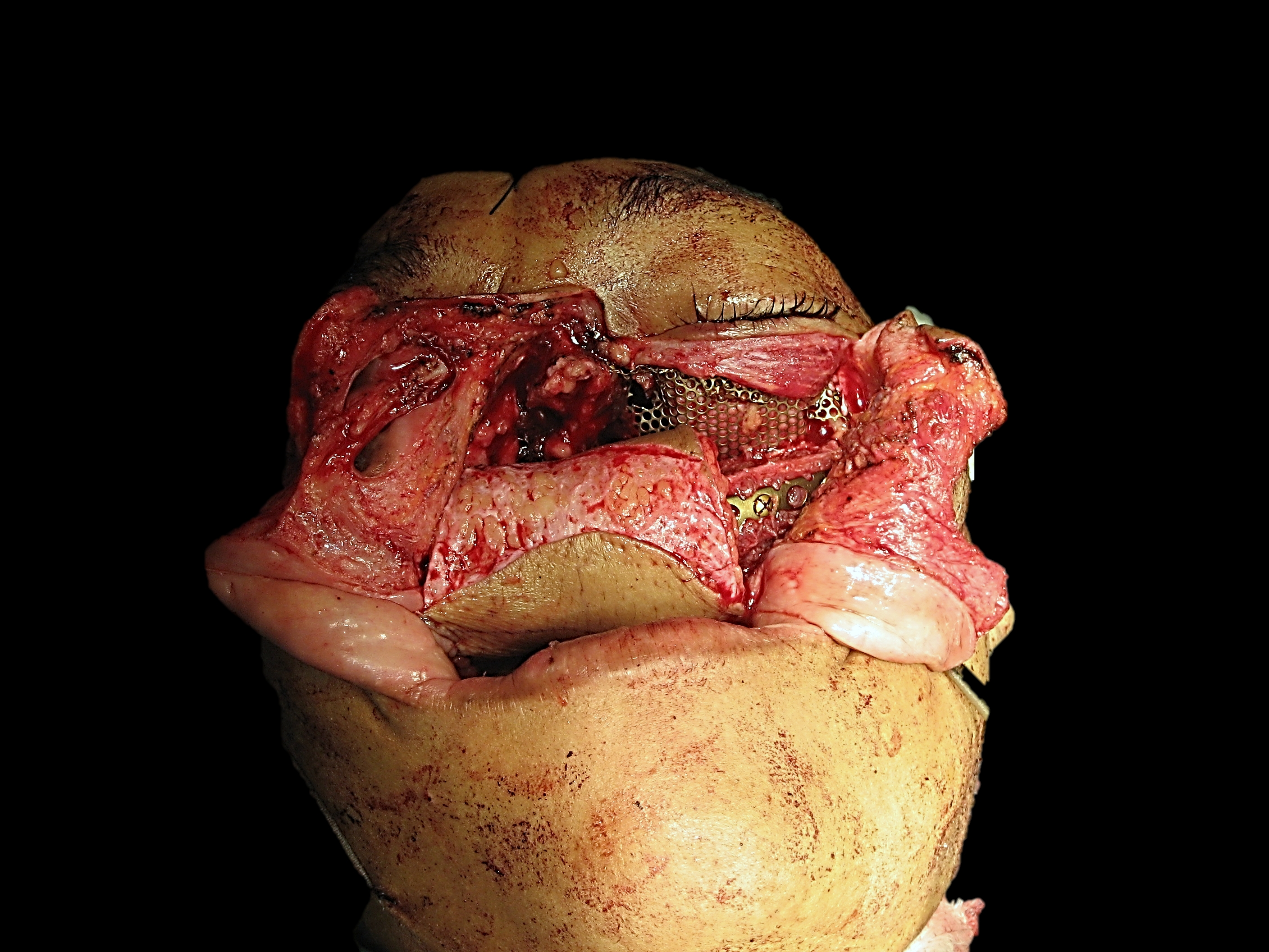
Enophthalmos, diplopia: Suture tears in the periorbita to avoid herniation of orbital fat. It is not unusual for patients to complain of some diplopia, but this usually improves with the passage of time. A degree of enophthalmos is to be expected unless the medial and inferior walls of the orbit are carefully reconstructed. If an extended resection of the orbital floor has been done, then consideration should be given to reconstructing the floor with fascia, bone or titanium mesh (Figure 44).
Figure 44: Free fibula flap following bilateral maxillectomy with reconstruction of orbit with titanium mesh
Extended total maxillectomy resections
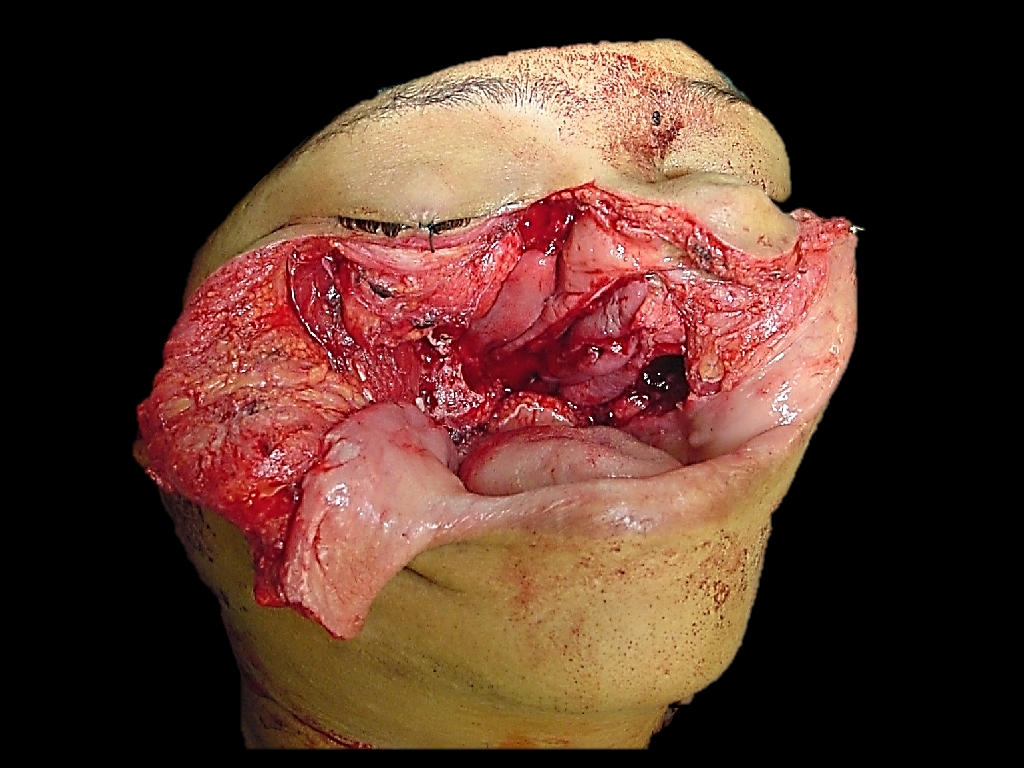
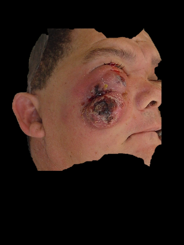
Total maxillectomy may be extended to include the malar complex, sphenoid sinus, cribriform plate (craniofacial resection), contralateral maxilla (Figure 45), skin (Figure 46a-c), or orbit. When resection extends to the contralateral maxilla (Figure 45), then an osseocutaneous free flap reconstruction (Figures 43, 44) is imperative.
Figure 45: Bilateral (total and inferior) maxillectomies
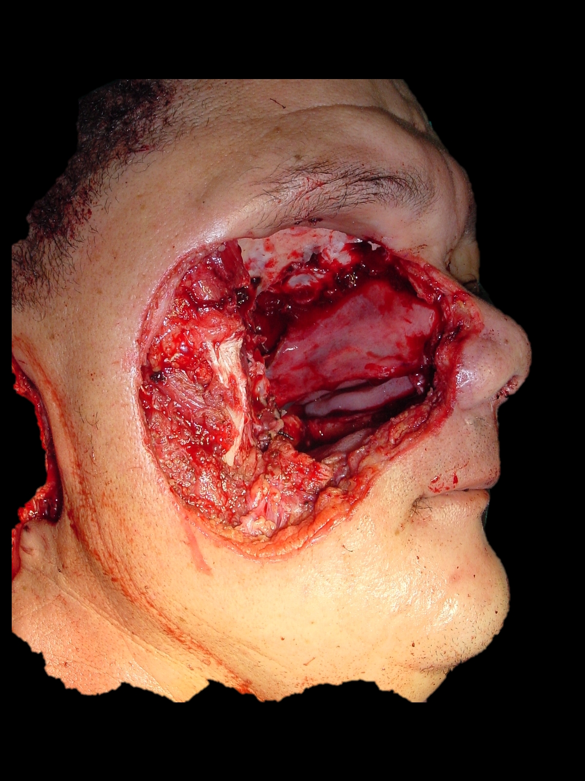
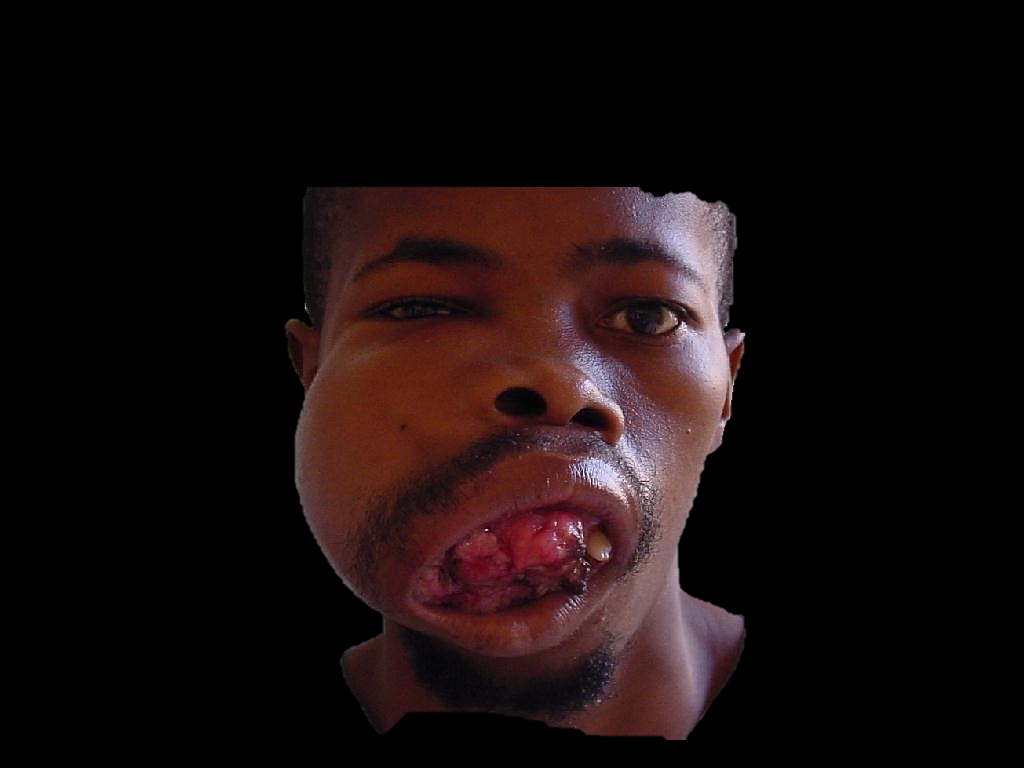
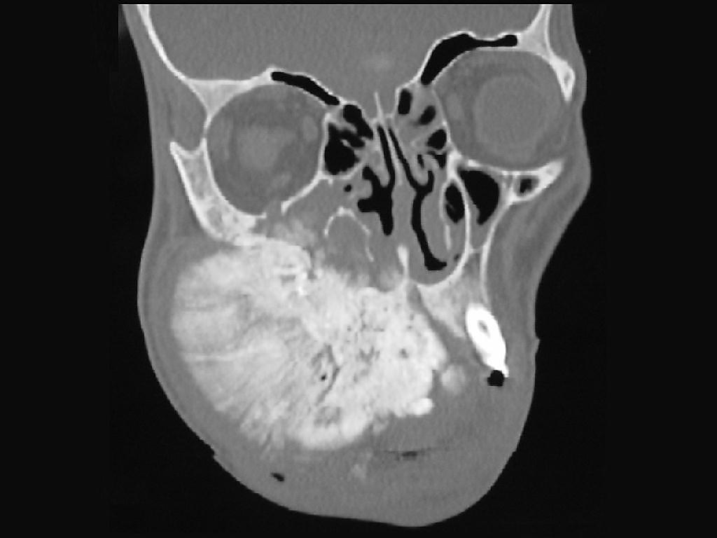
The patient depicted in Figures 46a-c required resection of overlying skin and soft tissues, infratemporal fossa and orbit, as well as nodal metastases to the parotid and neck. The defect was reconstructed with an anterolateral free thigh flap.
Figure 46a: Skin and orbital involvementFigure 46b: Orbital, infratemporal fossa and masticator space involvementFigure 46c: Total maxillectomy, orbital exenteration, resection facial skin, and parotidectomy
Orbital Exenteration
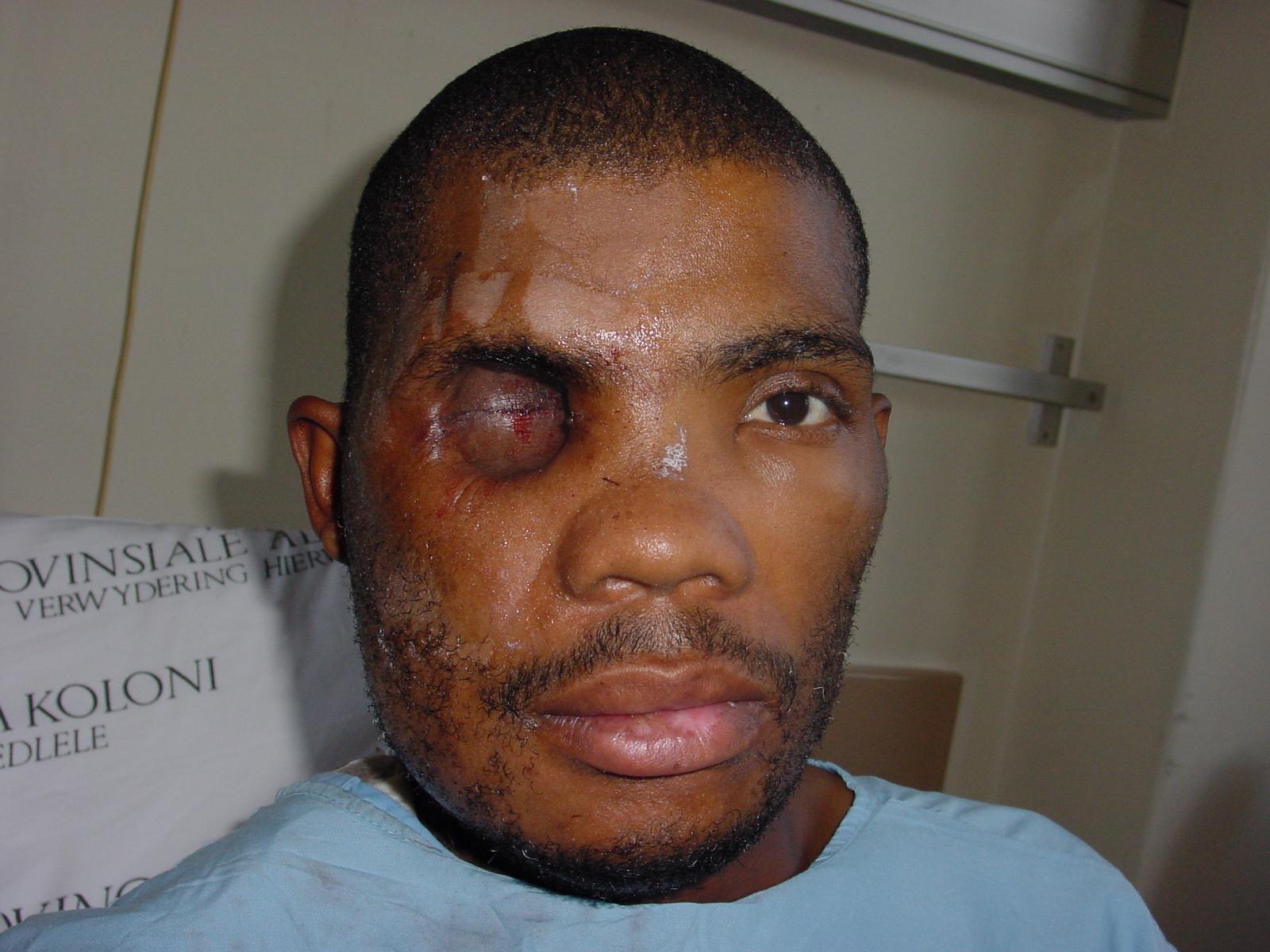
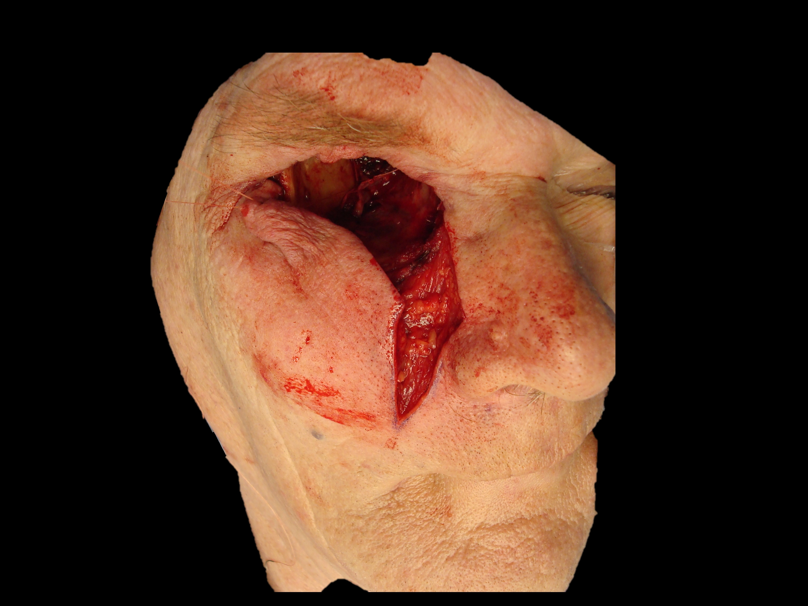
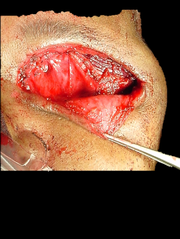
Orbital exenteration is generally indicated when tumour has extended through periosteum to involve fat (Figure 46, 47) and/or muscle and/or the globe; the eyelids; and the lacrimal apparatus. Involvement limited to the bone or periosteum can be managed by resecting the involved periosteum.
Figure 47: Tumour invading orbital fat
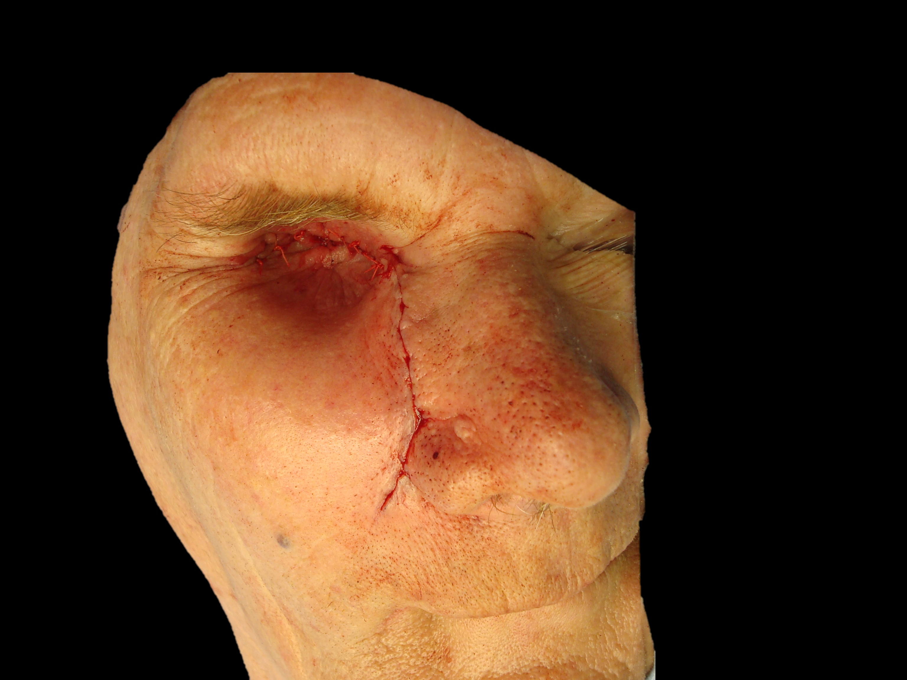
Whenever possible the eyelids are preserved (lid-sparing cf. lid-sacrificing exenteration) so they may be sutured together at completion of the surgery by making palpebral incisions just above and below the eyelashes. The skin is then elevated from the tarsal plates with monopolar cautery up to the orbital rims circumferentially.
The periosteum is incised circumferentially just medial to the orbital rim, exposing the underlying bone. Take care not to injure the supraorbital nerve so as to preserve sensation to the forehead.
The medial and inferior orbital dissections are as described on Page 9 of this chapter with transection of the lacrimal sac, and ligation and transection of the anterior and posterior ethmoidal arteries.
Next the orbital contents are mobilised in the relatively avascular subperiosteal plane inferiorly, laterally and superiorly. This is achieved by simply stripping the periosteum from the bone with e.g. a Freer’s dissector, other than along the inferior orbital fissure where the tissues are divided with scissors.
All that remains is to transect the ocular muscles and optic nerve and ophthalmic vessels at the orbital apex. Avoid undue traction on the optic nerve as this can injure the contralateral optic nerve. Exposure may be improved by decompressing the globe with a large bore needle. Use curved scissors (curved almost to 900) to transect the orbital apex and to deliver the orbital contents. Pack the orbit for a few minutes, and then use bipolar cautery or a ligature to control the brisk bleeding from the ophthalmic artery.
The orbital cavity may be managed in a number of ways. Patients may be left with the orbital cavity exposed +/- an (immobile) ocular prosthesis. When wearing a prosthesis is not an option, patients prefer to have the cavity filled with a flap.
Leave to granulate: This is a lengthy healing process
Split skin graft in the orbital cavity kept in place with gauze packing
Suturing eyelids together +/- a cheek advancement flap and inserting gauze packing to ensure that the skin is applied to the bone of the orbit as free floating skin will break down (Figure 48, 49a, b).
Figure 50: Temporalis muscle filling the orbit prior to suturing spared lower lid to superior orbital margin
Additional clinical examples
Figure 51a: Osteosarcoma of maxillaFigure 51b: Osteosarcoma of maxilla with expansion of inferior orbital canal indicating invasion of inferior orbital nerve; MRI is indicated to assess proximal perineural spread; resection should include malar complex and frozen section of inferior orbital nerve in pterygopalatine fossa
Author and Editor
Johan Fagan MBChB, FCORL, MMed
Professor and Chairman
Division of Otolaryngology
University of Cape Town
Cape Town
South Africa johannes.fagan@uct.ac.za