Cancers of the base of tongue (BOT) may be treated with primary surgery, and/or irradiation, and/or chemoradiation therapy. Both the oncology team and patient need to carefully weigh up morbidity vs. cure of surgical and nonsurgical options, both of which may cause significant morbidity. Patients need to be carefully assessed relating to their ability to deal with a measure of aspiration, access to speech and swallowing services and to PEG feeding should they not resume oral feeding.
Surgeons have to be au fait with the full range of surgical approaches and reconstructive options so as to ensure complete resection, minimise morbidity, and optimise speech and swallowing function.
Surgical approaches include the following:
This chapter focusses on surgical management of BOT cancer other than CO2 laser and TORS.
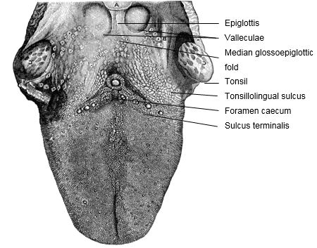
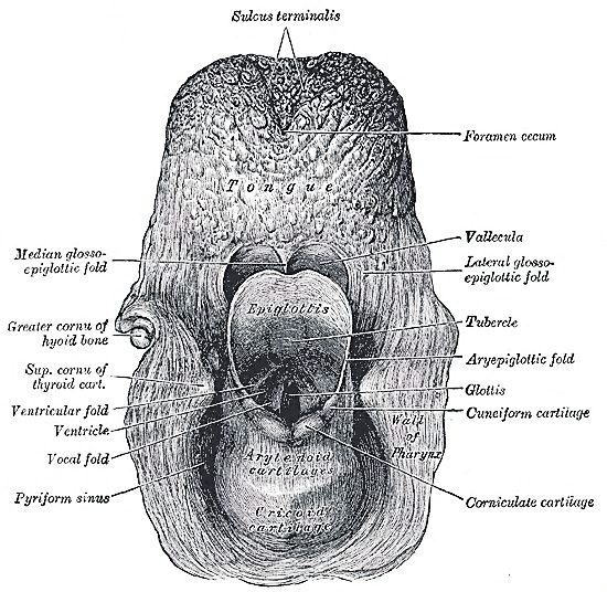
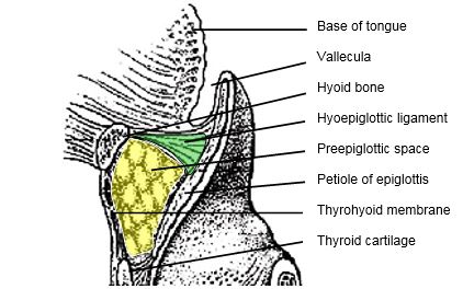
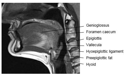
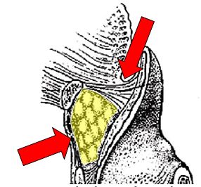
The BOT comprises the posterior 1/3 of the tongue behind the foramen caecum and sulcus terminalis (Figures 1, 2). The mucosa is rough, thick and fixed to the underlying muscle and contains lymphoid follicles (lingual tonsil); this makes it difficult to identify the edges of a BOT tumour; hence frozen section is especially useful to assess resection margins. Postero-laterally the tonsillolingual sulcus separates the tongue from the tonsil fossa. The valleculae separate the base of tongue from the lingual surface of the epiglottis and are divided in the midline by the median glosoepiglottic fold. (Figures 1, 2)
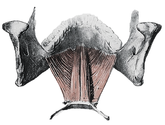
The tongue has eight muscles; four extrinsic muscles (genioglossus, hyoglossus, styloglossus, palatoglossus) control the position of the tongue and are attached to bone (Figure 3); four intrinsic muscles modulate the shape of the tongue, and are not attached to bone. Below the tongue are the geniohyoid and mylohoid muscles; the mylohyoid muscle serves as the diaphragm of the mouth and separates the tongue and FOM from the submental and submandi-bular triangles of the neck (Figures 3, 4).
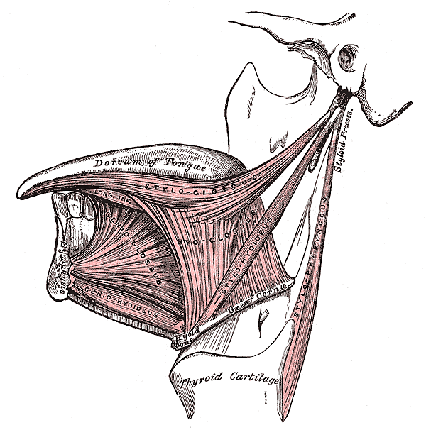
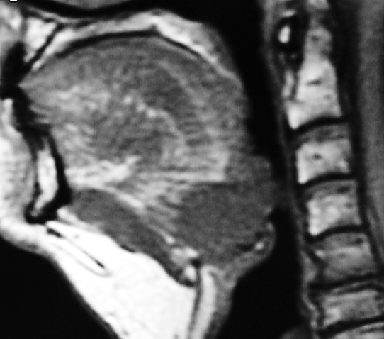
Posteriorly, the hyoid bone and preepiglottic space are important anatomical structures, an understanding of which is key to assessing whether the larynx can be preserved when using a suprahyoid surgical approach (Figures 5, 6). Most of the tongue muscles attach to the hyoid bone (Figures 3, 4); the lingual artery and hypoglossal nerve (XIIn) pass medial to the greater cornu of the hyoid (Figures 7, 8). The hyoid bone forms the anterior boundary of the preepiglottic space; the superior boundary is the hyoepiglottic ligament (floor of vallecula), and the posterior border is formed by the petiole of the epiglottis (Figures 5, 6).
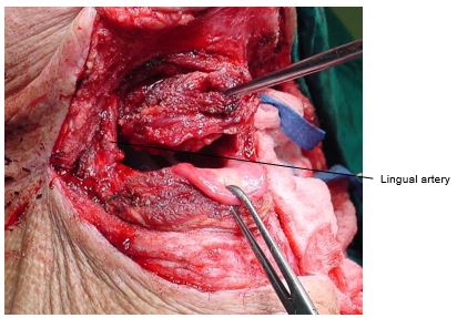
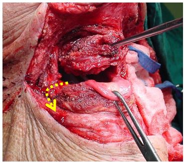
The arterial supply of the BOT is derived from the paired lingual arteries and its posterior (dorsalis linguae) branches (Figures 7, 8). Additional supply to the BOT emanates from the tonsillar branch of the facial artery and the ascending pharyngeal artery. In practice the lingual arteries are the only vessels that have to be looked for during BOT resection as they are easily injured; it is important to preserve at least one lingual artery to avoid infarction of the tongue. (The author has had one case where sacrifice of a single lingual artery led to necrosis of half the tongue; this is however unusual as there is usually adequate cross-flow).
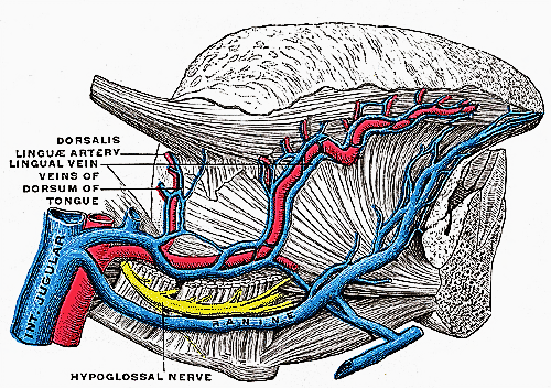
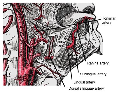
The lingual artery originates from the external carotid artery between the superior thyroid and facial arteries and courses obliquely forwards and medial to the greater cornu of the hyoid (Figures 7, 8). It then loops downward and anteriorly and passes medial to XIIn and the stylohyoid muscle. It then courses directly anteriorly and deep to the hyoglossus and finally ascends submucosally on the undersurface of the tongue as far as its tip as the ranine artery (profunda linguae); it lies to either side of the genioglossus, and is accompanied by the lingual nerve. Two or three small dorsalis linguæ arteries arise beneath the hyoglossus and ascend to the posterior part of the dorsum of the tongue and also supply the mucous membrane of the posterior floor of mouth (FOM) and oropharynx (Figures 7, 8).
Venous drainage is via lingual and ranine veins (Figure 7). The lingual veins originate on the dorsum, sides, and undersurface of the tongue and accompany the lingual artery and join the internal jugular vein. The ranine veins originate below the tip of the tongue and are visible on its ventral surface; they accompany the XIIn as venae comitantes and either join the lingual vein or pass lateral to hyoglossus to join the common facial vein.
An understanding of the nerve supply is important to preserve oral function. All intrinsic and extrinsic muscles are innerveted by the XIIn except for palatoglossus which is innervated by Xn. The IXn provides somatic afferent and taste sensation to the posterior 1/3 of the tongue. The lingual nerve provides general somatic sensation to the anterior 2/3 of the mouth and FOM; taste is provided by the chorda tympani branch of VIIn via the lingual nerve.
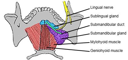
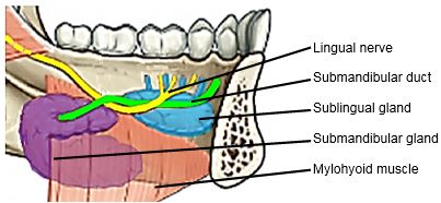
The lingual nerve is at risk when using a mandibular swing approach to resect tumours of the BOT. The nerve crosses deep to the submandibular duct in the lateral FOM; in the anterior FOM it is located posterior to the duct (Figures 9, 10).
The remainder of this chapter focuses on surgical resection of cancers of the BOT.
The surgeon always stands by the patient during induction of anaesthesia as it may be difficult or impossible to intubate a patient with a bulky BOT tumour that precludes elevation of the tongue to visualise the larynx. Should the anaesthetist be unable to intubate the larynx, the surgeon may be able to intubate with a laryngo-scope, or do an emergency tracheostomy or cricothyroidotomy; it is prudent to inject the tracheostomy or cricothyroidotomy site with local anaesthetic with adrenaline prior to induction. Nasal intubation facilitates resection of BOT tumours, and is conver-ted to a tracheostomy during the course of the operation.
Perioperative antibiotics are prescribed for 24hrs.
Good surgical access is essential in order to attain adequate resection margins, to control bleeding, and for reconstruction. A combination of open surgical approaches can be used, and will now be discussed. Level 1 of the neck should first be dissected if neck dissection is indicated before proceeding to address the primary tumour.
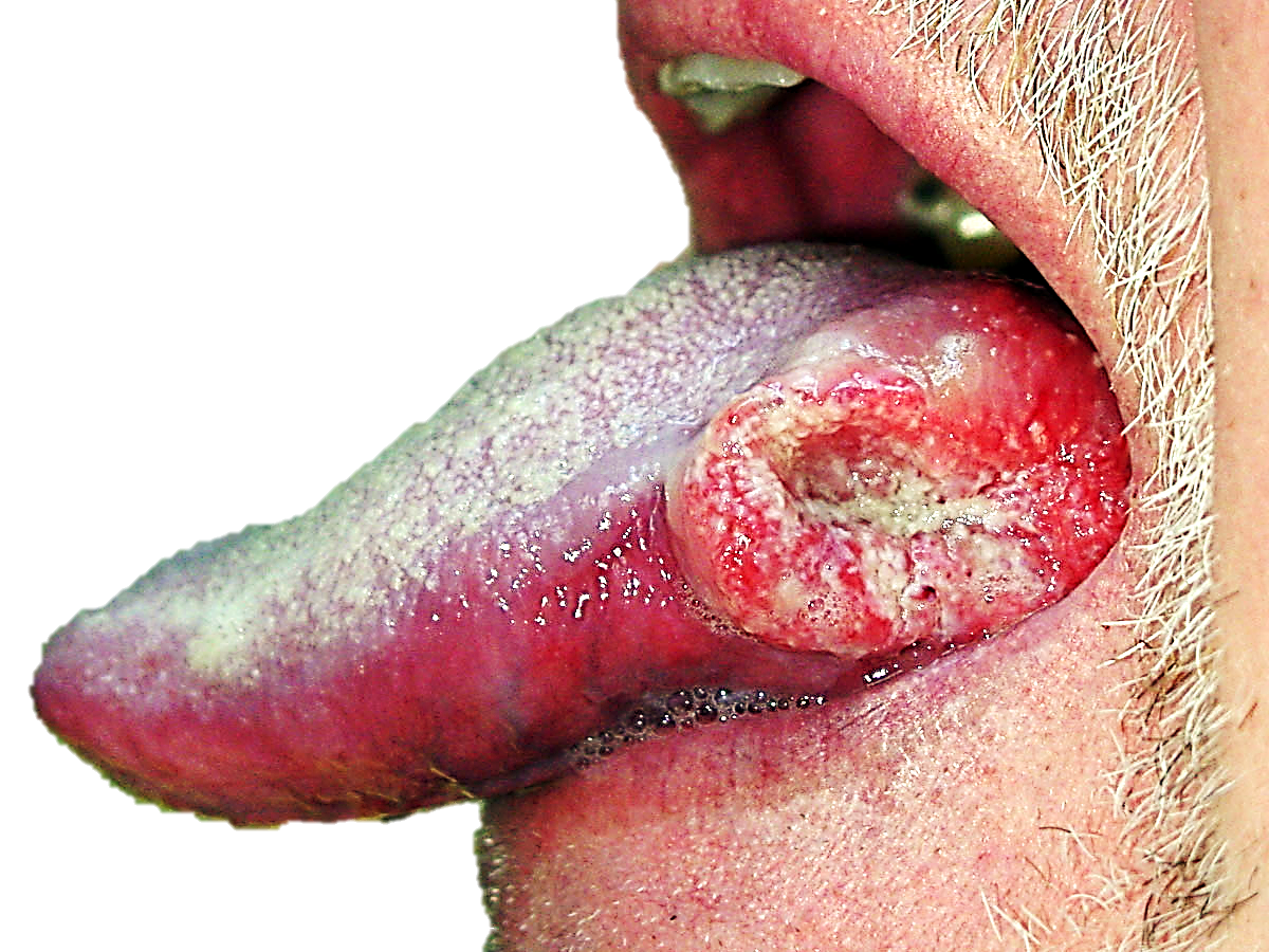
The adequacy of transoral access to the BOT varies considerably. A useful way to predict whether transoral resection is likely to be possible is to pull on the anterior tongue with a cotton swab during pre-operative clinical evaluation and to then see how accessible the tumour is. Note that tumours becomes more visible and accessible as the resection proceeds, especially once the thick BOT lining has been incised around the tumour. Laterally placed BOT cancers, especially in edentulous patients, are more amenable to trans-oral access (Figure 14).
The mouth is kept wide open either with a dental bite block (Figure 15) or with a self-retaining retractor, taking care to protect the teeth from injury (Figure 16). Apply traction to tongue and tumour with silk traction sutures or with tissue forceps (Figure 16).
Resect the tumour with at least a 1cm margin of normal tissue using electrocautery (Bovie). As the resection proceeds poste-riorly, insert new silk traction sutures or move the tissue forceps more posteriorly to facilitate delivery of the tumour into the mouth.
Midline BOT tumours may be exposed by bisecting the tongue along the median raphe with electrocautery (Figure 17); it is a relatively avascular plane as the vessels and nerves all course laterally, and results in little, if any, functional deficits. The incision can be carried posteriorly up to the hyoid bone if needed.

This affords excellent access to the BOT. It is especially well suited to cancers that extend anteriorly to involve the oral tongue (Figure 14), or onto the soft palate. It does however leave a facial scar; it may cause deformity of the lower lip; there is a risk of complications relating to the mandibulotomy and dental malocclusion; and the lingual nerve is at risk of injury.
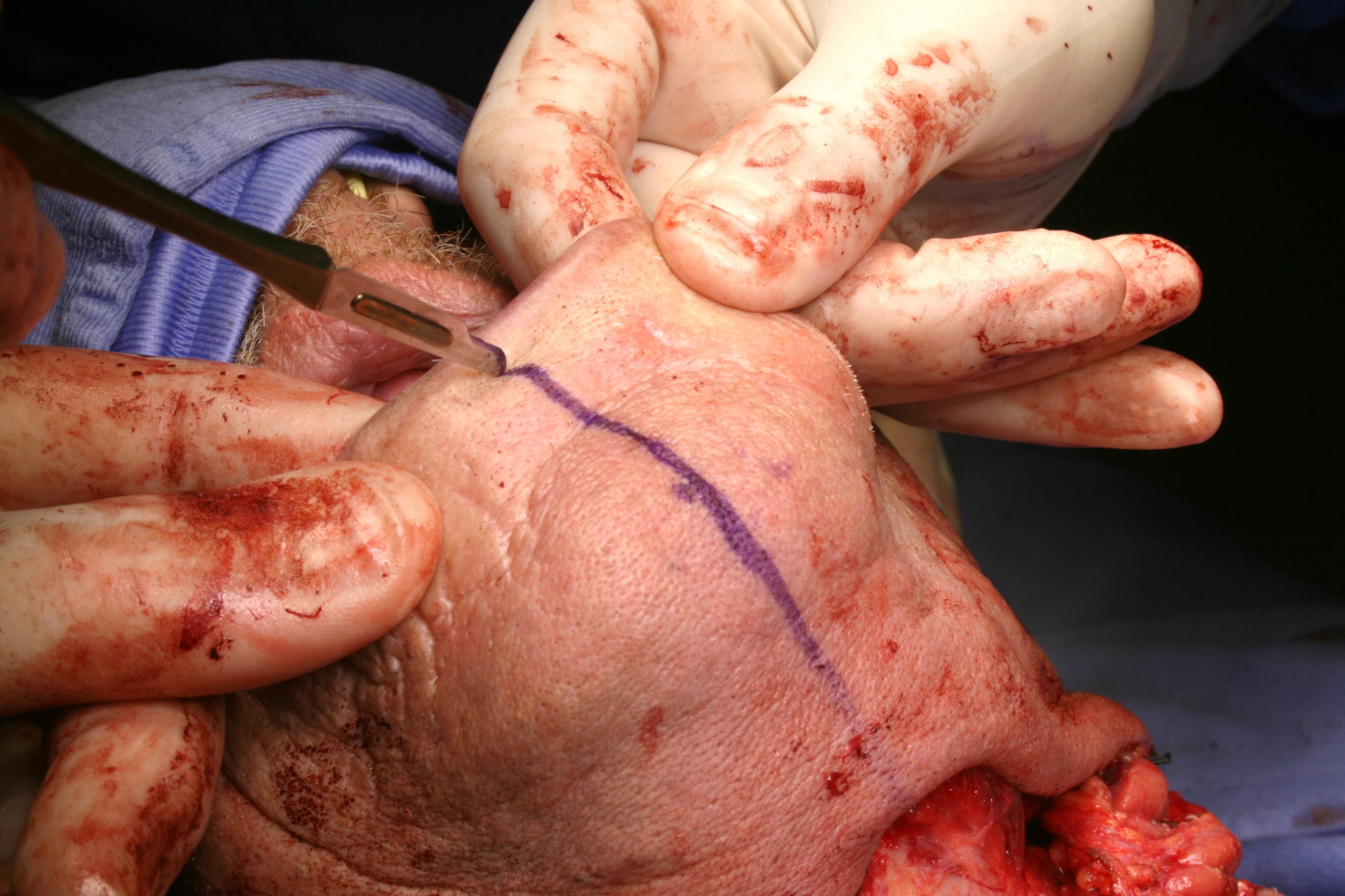
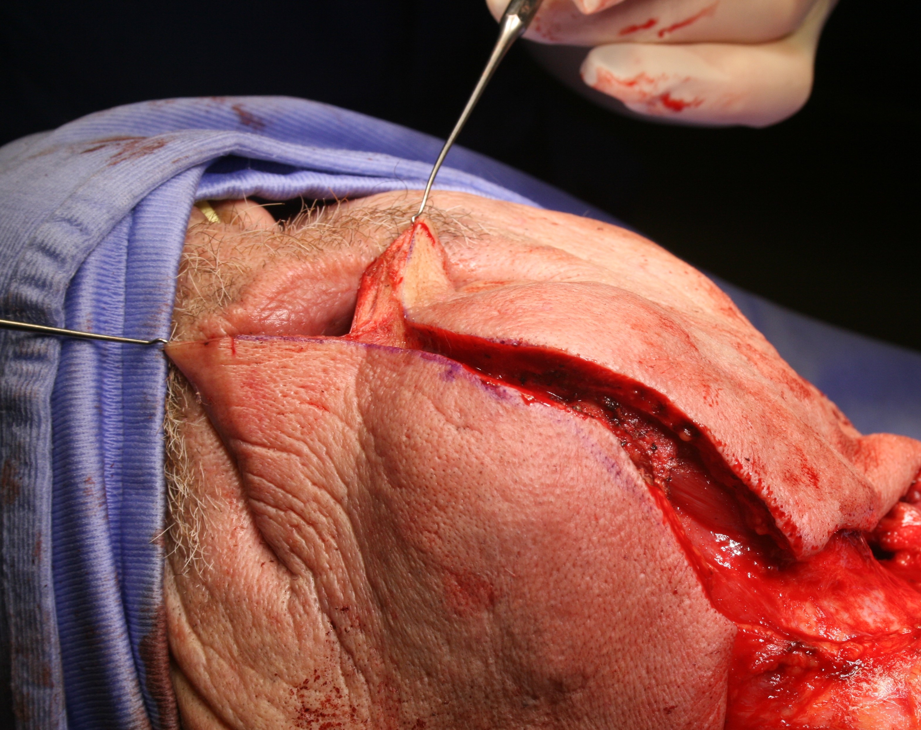
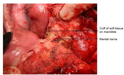
The vermillion border is scored or marked so as to ensure an accurate repair (Figure 18). The lower lip is divided vertically in the midline (Figure 19). Bleeding from the labial artery is controlled with cautery. Incise the gingivolabial and gingivobuccal mucosae >0.5cms from the bone leaving a cuff of soft tissue on the bone to facilitate subsequent soft tissue closure (Figure 20). Strip soft tissue off the mandible with monopolar cautery or with a periosteal elevator up to the mental foramen, taking care not to injure the mental nerve where it exits the foramen (Figure 20).
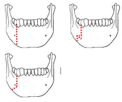
The mandible is divided just anterior to the mental foramen with a Gigli or a powered saw (Figure 21). The osteotomy may be made vertically or alternatively in a step- or V-shaped fashion so as to achieve a more stable repair (Figure 22). It is advisable to extract a tooth and make the osteotomy through the dental socket so as avoid devitalising adjacent teeth. In dentate patients the mandible is preplated with titanium miniplates contoured to the mandible so as to ensure perfect dental alignment. Two 4-holed, non-compression, 2mm mandibular plates, one placed along the inferior border of the mandible, and the other placed more superiorly are used. Once the plates have been contoured and the holes drilled, they are removed and the bone cut is made.
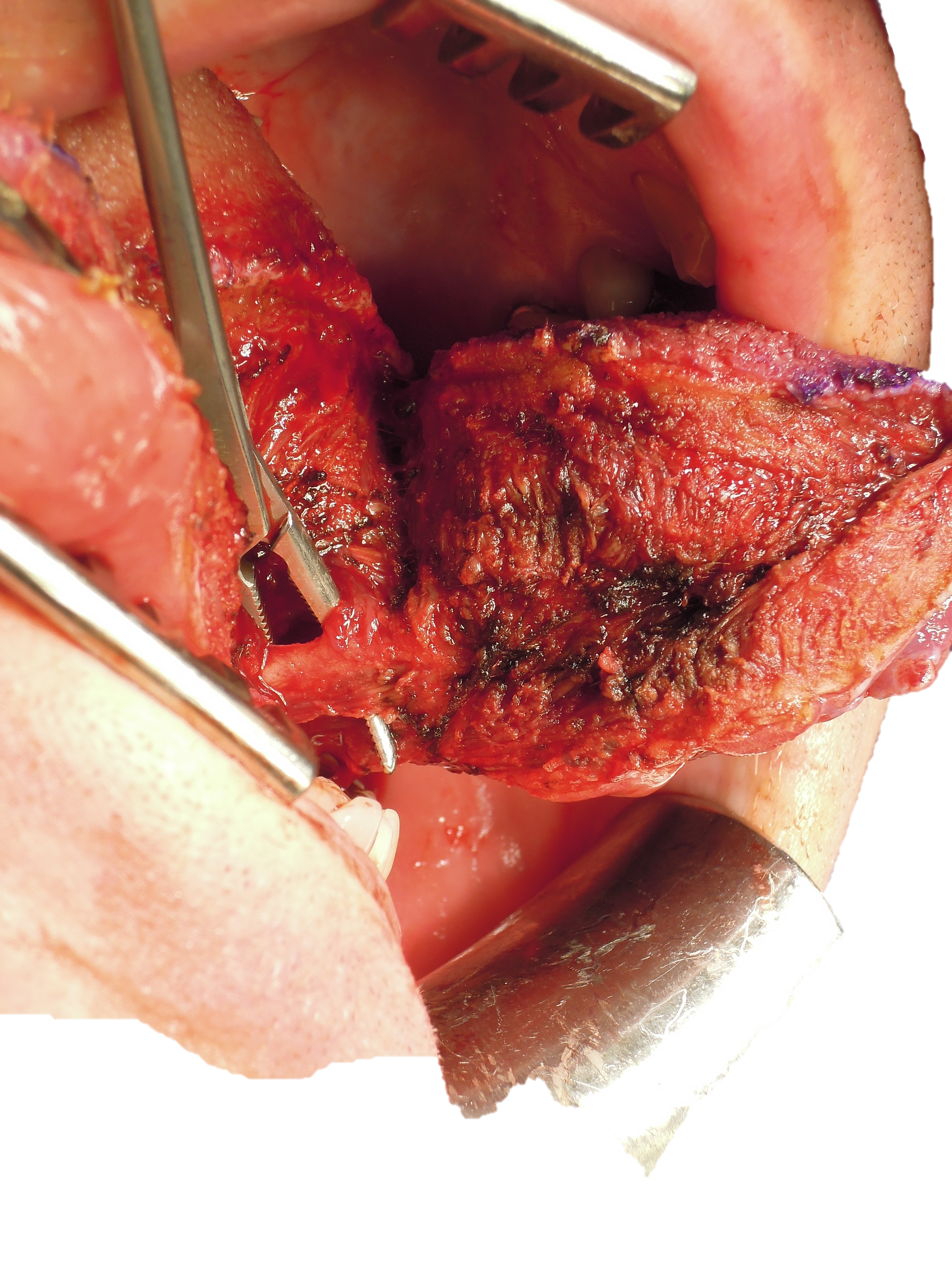
The surgeon then distracts the cut ends of the mandible to gain access to the oral cavity and proceeds to divide the floor of mouth (FOM) mucosa and mylohyoid muscle about 1cm from, and parallel to, the mandible, so as to leave soft tissue on the mandible for the subsequent FOM repair at conclusion of surgery. Continue the incision posteriorly along the FOM until the tumour comes into view; the lingual artery (medial to the hyoglossus muscle) and the XIIn course medial to the FOM incision are not at risk of injury at this stage of the dissection. The tumour is resected using electrocautery. The lingual artery may have to be ligated (Figure 23).
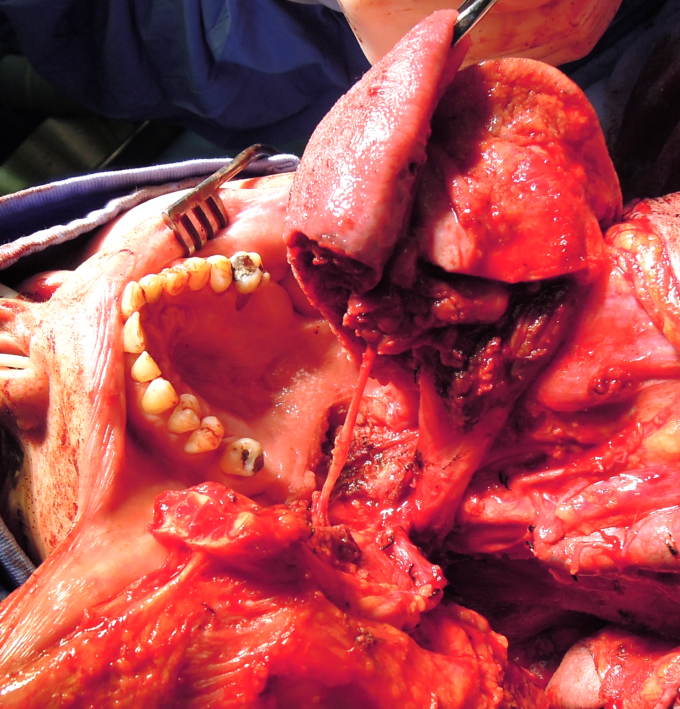
Posteriorly the lingual nerve extends from the skull base and crosses the line of the incision from lateral to medial to course along the lateral FOM; preserve the nerve if possible (Figure 24).
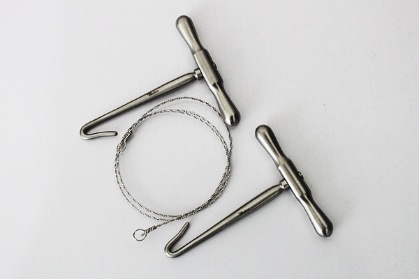
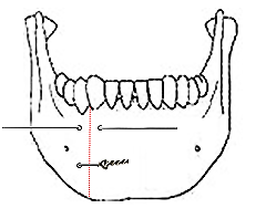
At conclusion of surgery the FOM is closed with a running vicryl suture, and the osteotomy is plated; when plating sets are not available, opposing holes are drilled on either side of the osteotomy and the mandible is wired together with stainless steel wiring (Figure 25). The lip is carefully repaired in layers to approximate the muscles as well as mucosa and skin.
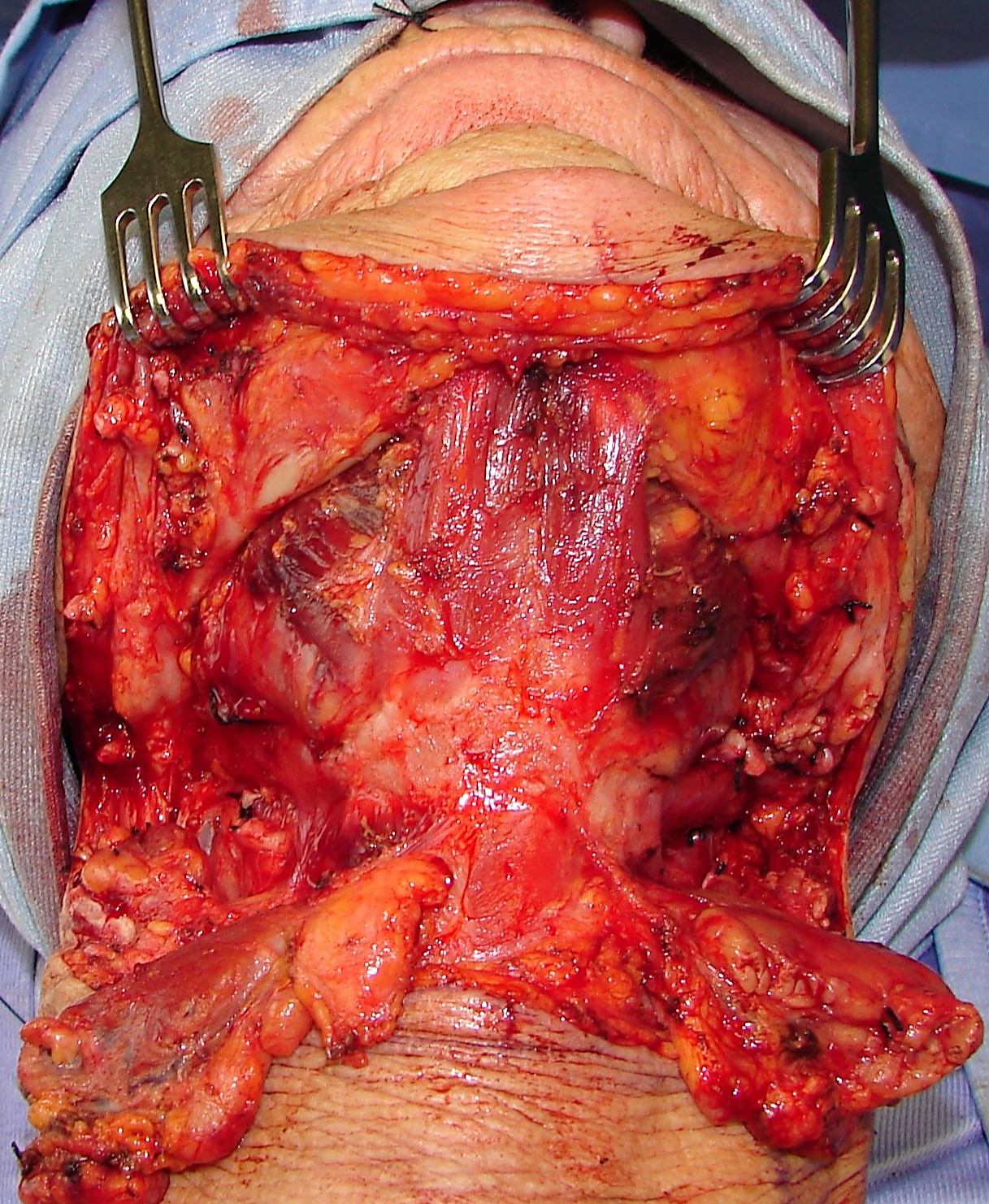
This is one of the external approaches favoured by the author. It is similar to the suprahyoid dissection step during total laryngectomy. It is well suited to most BOT cancers (Figure 26), although access is limited with cancers that extend far anteriorly into the tongue.
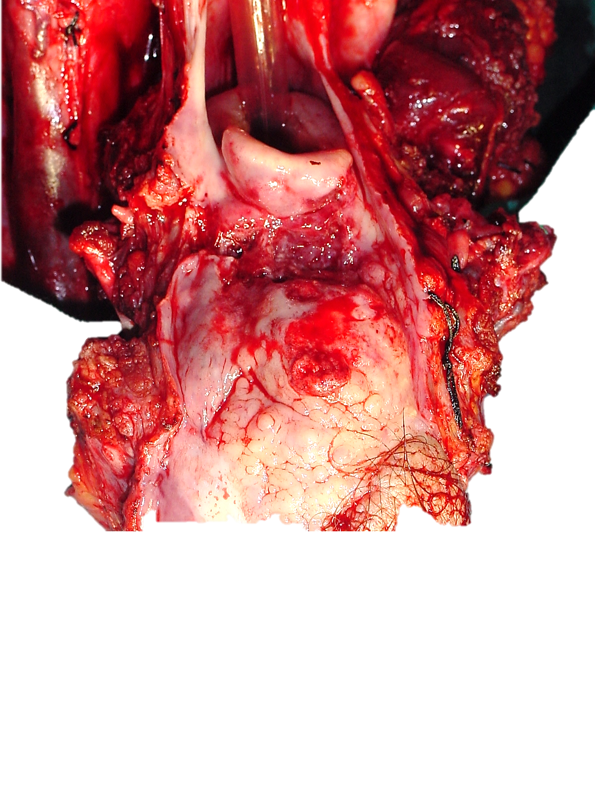
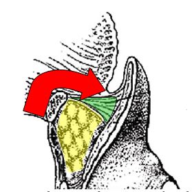
Because the pharynx is entered through the vallecula, it is not suited to cancers that involve the apex of the vallecula, preepi-glottic space or epiglottis (Figure 11). Following completion of Levels 1a and b of the neck dissection(s), the superior aspect of the body of the hyoid bone is skeleton-ised with electrocautery, keeping the dissection between the lesser cornua of the hyoid bone. The dissection is carried posteriorly above the hyoepiglottic ligament, which forms the roof of the preepiglottic fat (Figure 27). The vallecula is entered at its apex (Figure 27).
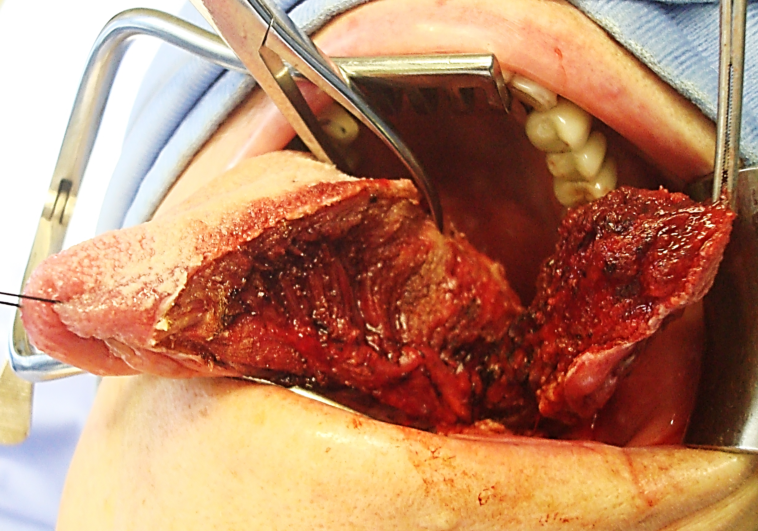
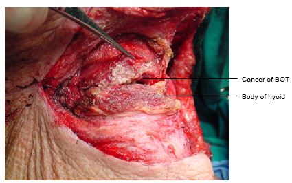
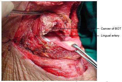
The tumour now comes into view. The cancer is resected with electrocautery (Figures 28-30). Take care to identify and preserve the lingual arteries and, if dissecting even more laterally, the XIIn (Figures 29, 30). Tumour exposure improves as the thick BOT lining is incised to either side of the tumour making the tumour more mobile (Figure 30).
Orientate the specimen for the pathologist with a suture before removing the specimen so as not to lose orientation. The adequacy of the resection margins is checked with frozen section if available.
Primary closure can almost always be done by approximating the BOT resection margin to the vallecula with a running vicryl suture. The supra- and infrahyoid muscles are then sutured together and the neck is closed in the usual fashion.
A temporary tracheostomy is done and a nasogastric feeding tube is inserted. Once the airway appears adequate the tracheostomy tube is corked for 24hrs before being removed.
Additional exposure can be obtained by extending the incision along the greater cornu of the hyoid bone and along the posterior margin of the thyroid cartilage; take special care to identify and not to injure the XIIn, the lingual artery or the superior laryngeal nerve (Figure 31).
This may be employed when tumour stops some distance from the inner aspect of the mandible. The mucosa of the lateral and anterior FOM is divided >0.5cm from the inner aspect of the mandible (so as to facilitate later repair), taking care not to injure the lingual and XIIns, or the submandibular ducts. Following bilateral neck dissections of Levels 1a and 1b, the mandibular attachments of the anterior bellies of digastric, mylohyoid, geniohyoids and genioglossus are divided with electrocautery working from inferiorly (Figure 32).This permits the surgeon to drop the FOM and tongue into the neck and to proceed with the resection. At conclusion of surgery the FOM mucosa and muscle are reapproximated with vicryl sutures.
Obtain meticulous haemostasis using ties, monopolar and bipolar cautery before closing the defect. Primary closure of BOT defects gives the best swallowing function although inadequate bulk impairs speech if the BOT cannot approximate the soft palate. Simply shaping a flap to match the resected tissue may well restore form but may have a poor functional result. Therefore one has to carefully assess the defect to determine how best to optimise swallowing and speech.
Radial free forearm flap: This is a thin and pliable flap and preserves mobility of the tongue, but has limited bulk.
Anterolateral free thigh flap: Muscle
harvested with the flap can be tailored to conform to the volume of the
defect to be filled. However it is less pliable than the radial free
forearm flap and is only suitable for oral reconstruction in patients
with thin thighs.
Pectoralis major flap: This is a good option to use (See chapter: Pectoralis major flap)
Buccinator flap: A posteriorly based buccinator flap can be used to close a small orocervical communication following resection of a lateral BOT cancer. (See chapter: Buccinator myomucosal flap)
Resecting tumours of the BOT is challenging particularly in terms of access and maintaining swallowing function. One should not compromise resection margins for function. The surgical team has to master an array of surgical approaches and reconstructive techniques so as to ensure the best oncological and functional outcomes.
Johan Fagan MBChB, FCORL, MMed
Professor and Chairman
Division of Otolaryngology
University of Cape Town
Cape Town, South Africa
johannes.fagan@uct.ac.za