MANAGEMENT PRINCIPLES/GUIDELINES FOR HEAD AND NECK CANCER IN DEVELOPING COUNTRIES
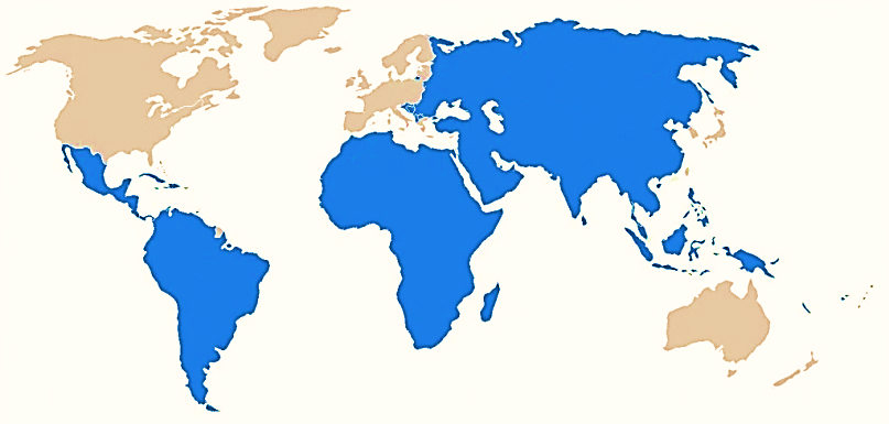
Developing countries constitute the majority of the world’s landmass (Figure 1) and are home to more than 50% of its people.
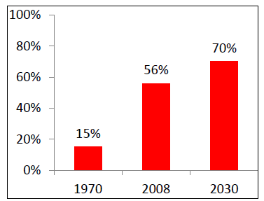
Cancer poses a major public health crisis in the developing world. Developing countries accounted for >50% of newly diagnosed cancers in 2010; it is projected that this figure will increase to 70% by 2030 (Figure 2). 1 This increasing percentage is attributed to population growth, reduced mortality from infectious diseases and an ageing society. 1
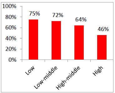
There is a wide disparity in cancer-related fatality which is aligned with income levels, ranging from 75% in low income countries, to 46% in high income countries (Figure 3). 1 Even though developing countries account for 67% of cancer-related deaths, they account for only 5% of cancer-related spending. 1
Therefore it is apparent that it is essential that innovation, expertise, resources, teaching and research be directed to addressing cancer in the developing world if we are to improve cancer outcomes globally. Given the financial and infrastructural challenges, the cancer management also has to be adapted to local constraints.
Head & neck cancer in developing countries
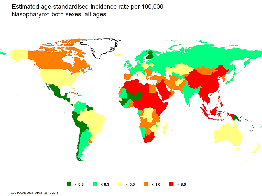
Rapid economic growth in developing countries has been accompanied by lifestyle changes associated with squamous cell cancer such as smoking, alcohol, and longevity. Two-thirds of oral and pharyngeal cancers (excluding nasopharynx) occur in developing countries. 2 Figure 4 illustrates the significant geographical variation for oral cancer; it is the most common cancer in males in high-risk areas such as Sri Lanka, India, Pakistan and Bangladesh where it accounts for up to 25% of all new cancers. 22 Cancer of the nasopharynx is also principally a developing world problem where it is related to Epstein Barr virus infection (Figure 5). 3,4,5
HIV is also associated with malignancies of the head and neck. The prevalence of HIV is highest in developing countries; sub-Saharan Africa accounts for two thirds of HIV positive people. 6 HIV is associated with Kaposi’s sarcoma and non-Hodgkin’s (and Hodgkin’s) lymphoma; with Kaposi’s sarcoma and lymphoma, surgery is restricted to obtaining a tissue biopsy. Squamous cell carcinoma of the conjunctiva is also associated with HIV. 7 Patients with advanced squamous cell carcinoma of the conjunctiva and metastases to parotid and cervical lymph nodes may require orbital exenteration, parotidectomy, neck dissecttion and orbital reconstructive procedures. The association between HIV and mucosal squamous cell carcinoma of the upper aerodigestive tract is less clear-cut. 8
Factors to consider when treating head & neck cancer in developing countries
One should guard against simply extrapolating management protocols from developed world centres to head and neck patients in developing countries.
-
Advanced cancers
Patients in developing countries are more likely to present with advanced cancer 9, 10; consequently treatment is mostly palliative 11. Even in a middle income country like South Africa, 52% of patients under-going total laryngectomy initially required an emergency tracheostomy 12 Late presentation may be due to ignorance, poverty, poor access to health services, and patients consulting traditional healers and using traditional medicines.
The adverse consequences of delayed presentation are compounded by long waiting lists for surgery and irradiation. Frequently patients become inoperable while awaiting surgery or radiation; this complicates initial patient selection and treatment planning. Jensen et al (2007) reported that one month’s delay was associated with a 62% increase in tumour size, 20% new nodal metastases, and that cancers were upstaged (TNM) in 16% of patients studied; mean tumour volume doubling time was 3 months. 13 Some institutions administer “holding chemotherapy” (methotrexate or platinum-based drugs) in an attempt to slow tumour progression while patients await definitive treatment; yet there is no evidence that this improves outcomes.
-
Does HIV status matter?
Do HIV +ve patients need to be managed differently? One may need to consider the following questions when managing HIV +ve patients, especially when resources are limited:
Is radiotherapy accompanied by increased mucosal and cutaneous toxicity in HIV +ve patients? Although there are many reports of radiotherapy-induced skin and mucosal toxicity with Kaposi sarcoma, the few reports of toxicity with other head and neck malignancies indicate good tolerance to radiation +/- chemotherapy.14, 15, 16
Should antiretroviral therapy be commenced to boost CD4 counts in immunocompromised patients prior to (chemo) radiation? Radiation can suppress CD4 counts. Yet even though it may seem reasonable to institute antiretroviral therapy to boost depressed CD4 counts prior to radiation, there are no controlled studies that address this question. Although interactions between antiretroviral therapy and radiation have not been well documented in the literature, there is a theoretical concern about the combination of the myelosuppressive effects of certain antiretroviral agents and myelosuppressive chemotherapeutic agents used with head and neck cancers such as platinum alkylators e.g. cisplatin and carboplatin. 16
What is the anticipated life expectancy of an HIV +ve patient? Adults that commence antiretroviral therapy before CD4 counts drop to < 200 cells/mm3 have about 80% of normal life expectancy; even severely ill HIV patients treated with antiretrovirals have at least an 80% chance of surviving 2 years. 17
Do CD4 count and HIV status affect surgical outcomes? Even major surgery does not depress CD4 counts 18, and HIV status per se does not increase the likelihood of early surgical complications. 19 A depressed CD4 count (<100 cells/mm3) has however been reported to be a predictor of postoperative sepsis. 20, 21 Instituting antiretroviral therapy prior to surgery has the benefits of reducing the viral load of the patient (viral exposure to surgical team), and increases patients’ CD4 counts.
It may be concluded that there is insufficient evidence to modify treatment of “apparently healthy” HIV +ve patients with head and neck cancer with CD4 counts of >350cells/mm3. 16 There is also little reason to routinely determine the HIV status of healthy-looking head and neck cancer patients from an oncologic perspective alone; only when HIV infection causes ill health and immunosuppresssion may HIV status preclude patients from undergoing major surgery or chemoradiation.
-
How does one prioritise head and neck cancer patients?
Deciding who or who not to treat when the burden of cancer exceeds available treatment resources is perhaps the most difficult task that oncologists and surgeons in developing countries have to deal with. Arriving at a decision involves ethical and practical considerations such as tumour stage, prognosis, palliation vs. cure, comorbidities, nutritional status, age, socioeconomic status, social support structure, distance from the closest treatment centre, likelihood of regular follow-up, parental status, employment status, and the difficult ethical issue of possibly denying access to publicly funded treatment to patients originating from a foreign country without adequate treatment facilities.
However, when access to surgery and radiotherapy are the principal treatment constraints, it is reasonable to prioritise patients with the most curable (early stage) malignancies, especially when adjuvant radiation is not available or will be significantly delayed following resection of advanced malignancies.
-
Radiotherapy
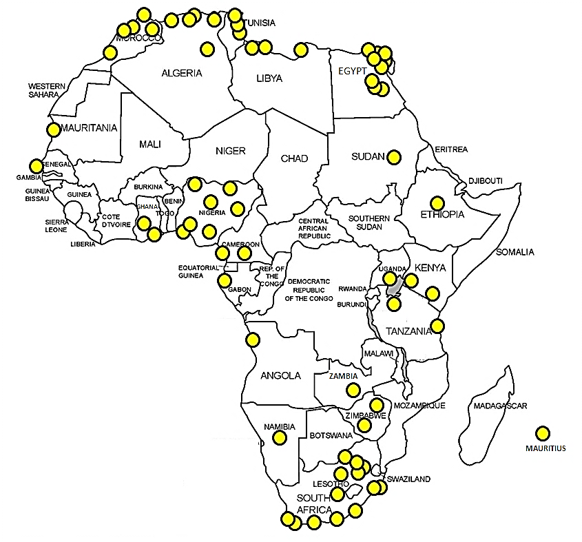
Though central to treatment of head and neck cancer, radiotherapy is unavailable in much of the developing world. Abdel-Wahab et al (2013) reported that only 23/52 African countries had radiotherapy facilities, and that facilities were concentrated in the southern and northern parts of the continent (Figure 6); that brachytherapy resources were available in only 20 countries; and that, because only 2% of African countries had modern imaging equipment and treatment planning systems, simple, curative treatment is generally based on two-dimensional imaging and treatment planning. 11
Tatsuzaki & Levin (2001) similarly reported significant unavailability of radiation facilities in Asia and the Pacific regions; 22 and Zubizarreta et al (2004) reported a major restriction to access to radiotherapy in 16/18 South American countries due to insufficient numbers of specialists. 23
Consequently most radiation services in the developing world are fairly basic and deliver mainly palliative care. 9 Radiation therapists also need to be cautious about extrapolating favourable treatment results emanating from modern radiation therapy centres of excellence to situations where reliance is placed on dated technology.
Patients that undergo radiation to the head and neck require long term follow-up to detect and manage delayed radiotherapy-related complications e.g. hypothyroidism increases over time and is present in 25% of patients at 5 years. 24 Therefore reliability for follow-up and the ability to monitor thyroid function and treat hypothyroidism have to be considered when selecting patients for radiation.
-
Chemoradiation
In developed countries, chemoradiation (mostly chemotherapy used concurrently with radiotherapy / CCRT) is widely used as an organ-sparing treatment strategy with squamous cell carcinoma of the oral cavity, larynx, and oro-, hypo-, and nasopharynx. Compared to radiotherapy alone it has an 8% advantage in terms of locoregional control and survival rates. 25
However, to achieve such favourable outcomes, the “package of care” must include modern, sophisticated imaging (CT, MRI, PET) both for treatment planning and follow-up, medical and intensive care support for chemotoxicity, PEG feeding, and complex salvage surgery for persistent cancer or for recurrence as well as dental, speech, swallowing and audiological rehabilitation. Salvage surgery requires high levels of surgical expertise including proficiency with free tissue transfer flaps.
Because chemoradiation is expensive, toxic 25 and complex treatment and requires a “package of care” not available in many developing world centres, it has to be employed with great circumspection in a developing world setting.
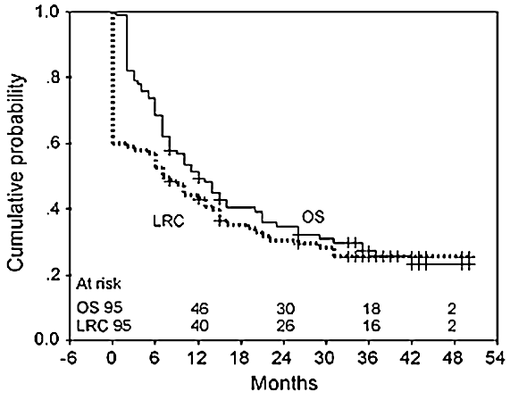
Kumar et al. reported a 14% mortality rate during and within 30 days of treatment in patients with advanced head and neck cancer treated with concomitant boost radiotherapy with concurrent weekly cisplatin at a tertiary hospital in India; the authors attributed the high mortality to poor support to deal with acute morbidity, poverty, malnutrition, illiteracy, and poor hygiene and concluded that “on present evidence in the setting of a developing country, CCRT with concurrent cisplatin cannot be recommended as primary therapy in advanced head and neck cancers without formal comparison with other treatment modalities” (Figure 7). 26
Therefore, if chemoradiation is to be considered, patients must be carefully selected to predict favourable outcomes by considering factors such as age, general health, social support, immune (HIV) status, tuberculosis, and the “package of care“ mentioned above should be available.
Altered fractionation
Altered fractionation schedules also improve locoregional control. Accelerated radiotherapy is perhaps better suited to a developing world setting than chemoradiation as it is cheaper and better tolerated. Overgaard et al reported that a 6-fractions-per-week radiation schedule significantly improved locoregional control for squamous cell carcinoma of the larynx, pharynx and oral cavity when compared to conventional schedules of 5-treatments-per-week. Despite increased acute morbidity, accelerated radiotherapy did not cause increased late morbidity and had the benefit of reducing overall treatment by 1 week. 27 Concomitant boost radiotherapy with a 2nd daily fraction to the gross tumour volume in the final 10 days of treatment also reduces overall treatment time, thus reducing the chance of repopulation and improves local control. 28
However unless a radiation therapy department already treats patients 6 days/week, both the above schedules require reorganisation to accommodate the 2nd daily fraction.
-
Surgery
Surgery is often the only treatment available because of inadequate radiotherapy and chemoradiation facilities. Yet, surgeons in developing countries frequently lack head and neck surgical training, and modern surgical technology (bipolar cautery, laser, transoral robotic surgery, endoscopic surgery), frozen section, blood products, adequate operating time, good anaesthesia and intensive care support are often lacking. 29
Surgeons in developing countries need to keep abreast of and adapt modern surgical principles and techniques to a lower technology type practice e.g. substitute trans-oral microsurgery for early laryngeal cancer with laryngofissure and other open partial laryngectomy procedures; ensure wide tumour resection margins in the absence of frozen section control and post-operative radiation therapy; liberally employ elective neck dissection in the absence of sophisticated imaging, and rely on a range of pedicled rather than microvascular free tissue transfer flaps to reconstruct surgical defects.
Management protocols
Selecting appropriate treatment for head and neck cancer patients in developing countries is particularly challenging and involves complex, individualised decisions often without the benefit of special investigations such as FNAC, CT, MRI, PET-CT and HPV status.
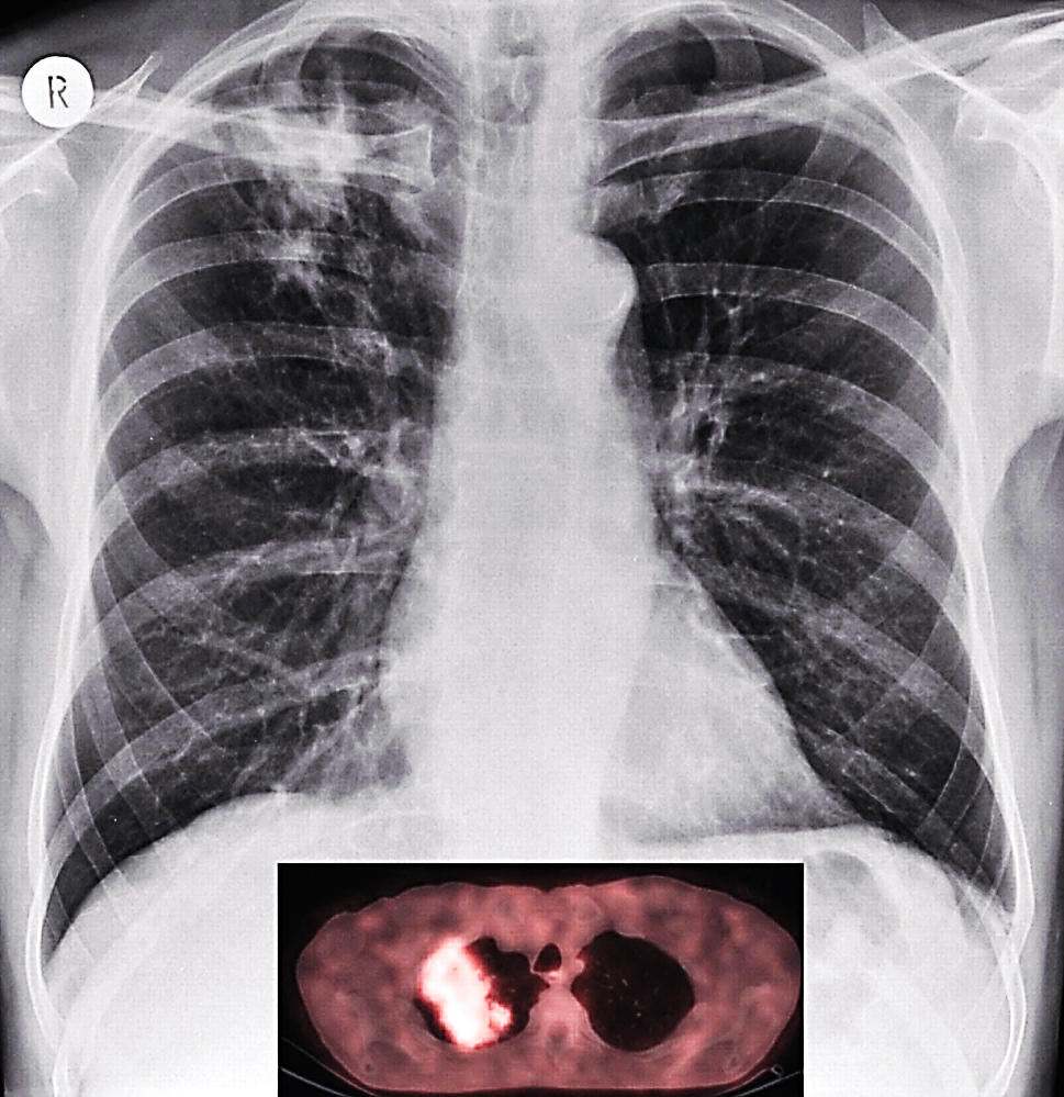
Unlike the situation in well-resourced health systems, it may not always be possible for treatment to be protocol-driven as the majority of patients are dependent on state run services with poor health infrastructure and resources. For the same reasons protocols designed for developed world settings are not always relevant; e.g.tuberculosis mimics metastases on PET scan, therefore limiting its utility as a staging tool in societies where tuberculosis is endemic (Figure 8).
Reliance therefore frequently is placed on clinical acumen, experience, intuition and institutional bias, often in the absence of scientific evidence to support clinical decisions. Investigations and treatment have to be tailored to the individual patient taking into account resource constraints relating to e.g. CT, MRI, operating rooms, ICU, radiation facilities, and blood transfusions; treatment delays (often many months);
likelihood of regular follow-up; access to drugs e.g. thyroid and calcium replacement; nutritional status; social support; poverty; comorbidities (often poorly treated or neglected) including HIV; cultural bias; and the availability of surgical expertise, radiation therapy and chemotherapy.
Certain principles will now be highlighted that may be considered when designing management protocols in resource constrained settings.
History: Consider cultural and religious values of patients as these may affect management. Inquire about risk factors including betel nut, areca nut, reverse smoking, chewing tobacco and comorbidities e.g. tuberculosis.
Metastatic workup: When access to operating time and adjuvant radiotherapy is limited, one could argue in favour of employing CT (even if it is a limited and expensive resource) to rule out pulmonary metastases that are not evident on CXR before (inappropriately) committing scarce surgical resources to advanced T and N stage cancers that have metastases.
N0 neck: It is reasonable to have a low threshold to electively treat the N0 neck by means of a selective neck dissection in cases of unreliable follow-up, lack of specialised imaging (initially and for follow-up), and delayed adjuvant radiation. Surgeons should have a low threshold to convert a selective to a modified neck dissection when suspicious lymph nodes are encountered, especially in the absence of postoperative radiation capability.
N+ neck: Even though lymphadenopathy in patients from poorer communities may result from untreated dental, oral and pharyngeal infections, HIV, or TB, palpable nodes within the expected lymphatic drainage area of a cancer should be treated with modified or radical neck dissection to avoid undertreating a neck that harbours metastases.
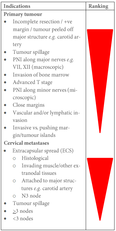
Adjuvant irradiation: When the burden of disease outstrips a centre’s capacity to provide postoperative radiation to all deserving patients, an oncologist may have to make a call as to who are most likely to benefit. Even though studies do not rank accepted indications for postoperative radiation, Table 1 is an attempted ranking (“thumb-suck”) of indications for adjuvant radiation (not to be cited). E.g. having ≥2 cervical metastases is generally considered to be an indication for adjuvant radiation, even though the evidence to support this threshold is tenuous; hence centres that lack capacity to provide radiation to all deserving patients could argue that this threshold be adjusted upwards so that patients most likely to benefit e.g. with positive margins, extracapsular spread (ECS), and large tumour volumes are not deprived of adjuvant radiation. 30 A final (major) caveat relating to using histological criteria for adjuvant radiation is the inaccuracy of histopathological (under)reporting, especially of the presence of cervical micrometastases and perineural invasion (PNI).
Reconstruction: Although excellent functional results can be achieved with micro-vascular free tissue transfer flaps, 31 the surgery is time-consuming and requires specialised training. In the absence of microvascular free tissues transfer flaps, surgeons should become proficient at using a range of pedicled flaps e.g. pectoralis major, buccinator, temporalis, nasolabial, buccal fat pad, deltopectoral, latissimus dorsi and forehead flaps.
Oral cavity: When postoperative radiation is unavailable, surgery for T1 and T2 cancers should be prioritised, including cancers that are T4 due to only limited bony invasion that can be resected by marginal or segmental mandibulectomy or by partial maxillectomy. Cancers of the tongue and floor of mouth that are palpable (likely to be >4mm thick) or are staged ≥T2 should undergo elective neck dissection due to the likelihood of there being occult cervical metastases. Preserving oral function is crucial; other than microvascular free transfer flaps, surgeons can employ pedicled flaps e.g. pectoralis major, buccinator, buccal fat pad, temporalis, nasolabial flaps. With inferior or total maxillectomy, one must separate the oral cavity from the nose; if prosthetic appliances are not available, this can be achieved with temporalis muscle flaps. Without the facility to reconstruct bone (e.g. free fibula flap), mandibular resection should not be extended beyond the midline so as to avoid the crippling and unsightly Andy Gump deformity (Figure 9).

Oropharynx: Management of cancers of the oropharynx has undergone a paradigm shift with the realisation that HPV infection is both an aetiologic and prognostic factor for a subset of oropharyngeal squamous cell carcinomas; the introduction of transoral robotic surgery (TORS) to resect oropharyngeal tumours as well as attempts to reduce the morbidity of chemoradiation by accepting smaller resection margins when combined with postoperative radiation. However HPV testing, transoral robotic surgery, and chemoradiation are generally not available in developing world centres; neither is the ability to deal with adverse consequences of chemoradiation. Surgical resection and/or radiation are therefore the mainstay of treatment in developing countries. Pedicled flaps used to reconstruct soft palate, lateral pharyngeal wall or base of tongue include pectoralis major, buccinator, buccal fat pad, and temporalis flaps.
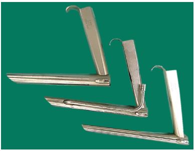
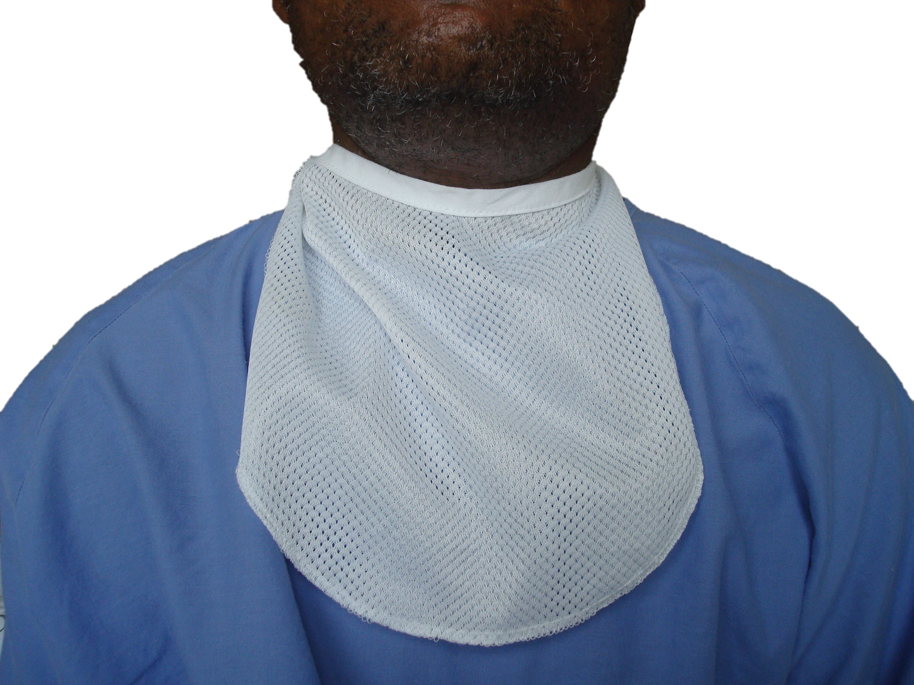
Larynx and Hypopharynx: In developed world centres early cancers are commonly excised with CO2 laser. Advanced cancers (dysfunctional larynx, cartilage invasion, tracheostomy for stridor) are treated with total laryngectomy. The remainder are offered chemoradiation with surgery reserved for salvage for persistent or recurrent cancer. CO2 laser is generally not available in developing world centres; chemoradiation is expensive and the package of care required to manage both acute and late consequences and complications of chemoradiation (dysphagia, PEG feeds, cancer surveillance with MRI and PET scans, complex salvage surgery, hypothyroidism, hypocalcaemia) is lacking. Therefore such centres have to rely on open approaches such as laryngofissure, vertical partial, supraglottic, supracricoid and near-total laryngectomy for smaller cancers, and total laryngectomy for advanced cancers. When performing total laryngectomy the surgeon should attempt to preserve both thyroid lobes and the parathyroids to minimise the risks of hypothyroidism and hypoparathyroidism, particularly when thyroid and calcium monitoring and replacement are difficult or impossible. With a dedicated speech therapy service, fistula (Figure 10) speech results can be achieved that match those of developed world centres even with poor, illiterate patients living long distances from treatment centres. 32 Voice prostheses are however expensive; hence the adoption of strategies such as using removable prostheses as indwelling prostheses to reduce expense 32. Heat moisture exchange (HME) devices are used in developed world centres to humidify and warm inspired air; however a homemade cloth stoma cover/bib (Figure 11) is equally effective at a fraction of the cost (Quail et al; unpublished study).
Although oesophageal speech does not cost anything, only 27% of patients in a Brazilian study mastered oesophageal speech 33. Another option is to use mucosal shunts; however the surgery is technically difficult and can be used only in highly selected patients with good pulmonary function who can cope with aspiration. 33 Because there is a severe shortage of speech therapists in many developing countries 28, 33 an electrolarynx is a reasonable alternative to achieve postlaryngectomy speech.
Nasopharynx: Cancers of the nasopharynx occur mainly in the developing world (Figure 5). Chemoradiation is the mainstay of treatment. 34 However the supportive care required for the extreme chemotoxicity is not always available in developing countries. Patients generally present with advanced disease; in an unpublished study conducted in Cape Town, South Africa, 50% of patients presented with Stage 4B disease of which 28% did not complete treatment for socioeconomic reasons (Dalvie et al: unpublished data). Hence it may be prudent to accept lower survival using radiation alone, than to try to improve survival with concurrent CTRT with its attendant morbidity. Intensity modulated radiotherapy (complex 3D conformal radiotherapy) has a documented survival benefit and can improve quality of life due to reduced xerostomia; however this generally is not available in developing countries. 34
Thyroid: Most thyroidectomies in developing countries are done by surgeons not specialising in endocrine surgery. Bilateral recurrent laryngeal nerve injury causing airway compromise, or hypoparathyroidism causing hypocalcaemia in situations where monitoring serum calcium and treating hypocalcaemia with calcium and Vitamin D are not possible may have fatal consequences. Regardless of surgical expertise, complication rates rise with the extent of resection. Subtotal thyroidectomy preserves the blood supply to the parathyroid glands and reduces the risk of hypocalcaemia. Thyroid lobectomy almost never causes significant hypoparathyroidism. Total thyroidectomy is however associated with both increased short- and long-term morbidity relating to recurrent laryngeal nerve paralysis and hypocalcaemia, particularly in an occasional thyroid surgeon’s hands. In the absence of convincing evidence that total thyroidectomy confers survival benefit in favourable, differentiated thyroid cancer 35, 36 (especially when I131 therapy is not available), coupled with the morbidity and mortality of total thyroidectomy where calcium monitoring and replacement are suboptimal, the occasional thyroid surgeon practising in a developing world centre may be wise to perform thyroid lobectomy or subtotal thyroidectomy for such cases.
Prevention and Screening
Given the cancer tsunami that the develop-ping world is facing, and the late presentations of patients with advanced disease, prevention (education and anti-smoking campaigns) and screening would appear to be reasonable strategies to adopt. Yet a Cochrane review reported that visual screening for oral cancer did not have survival benefit, although there was some evidence that it might be effective in high-risk patients. 37, 38 Techniques using toluidine blue staining, brush biopsy or cytology, or fluorescence imaging as primary screening tool or as adjunct for screening have also not been shown to have any benefit 37,. Consequently it appears that based on current evidence, there are more important interventions than investing scarce human and financial resources in screening that will improve head and neck cancer outcomes in a resource constrained setting. Our efforts should rather be directed towards educating patients to seek advice early when symptoms appear, educating doctors and health care workers to recognise potential malignancies and to refer patients early for appropriate management.
Concluding Remarks
There needs to be a global effort to educate and train oncologists and surgeons to manage head and neck cancer in developing countries through residency programmes, clinical fellowships and outreach projects. These should focus on developing sustainable head and neck cancer programmes, integrating them with existing local services and focusing on teaching and training. Open access to journals and text-books should be encouraged. In addition, a multifaceted approach is required including lobbying international organisations, governments and aid organisations to support infrastructure development and research, and for industry to provide appropriate and affordable technology. In this way the developed world can make a substantial difference to the outcome of the enormous head and neck cancer burden in the developing world.
References
- Farmer P, Frenk J, Knaul FM, et al. Expansion of cancer care and control in countries of low and middle income: a call to action. Lancet. 2010 Oct 2;376 (9747):1186-93
- Warnakulasuriya S. Global epidemiology of oral and oropharyngeal cancer. Oral Oncology. 2009;45:309-16
- GLOBOCAN 2008: International Agency for Research on Cancer (IARC), 2013.
- Yoshizaki T, Ito M, Murono S, et al. Current understanding and management of nasopharyngeal carcinoma. Auris Nasus Larynx 2012;39:137-44
- Maxwell JH, Kumar B, Feng F, et al. HPV positive, P16 positive, EBV negative nasopharyngeal cancer in White Americans. Head Neck 2010; 32(5): 562-7
- UNAIDS 2012 http://www.unaids.org/en/regionscountries/countries/southafrica/ (Accessed on 19 October 2013)
- R Newton, J Ziegler, C Ateenyi-Agaba, et al. The epidemiology of conjunctival squamous cell carcinoma in Uganda. Br J Cancer. 2002 July 29; 87(3): 301–8
- Engsing FN, Gerstoft J, Kronborg G, et al. Head and neck cancer in HIV patients and their parents: a Danish cohort study. Clin Epidemiol. 2011; 3: 217–27
- Onyango JF, Macharia IM. Delays in diagnosis, referral and management of head and neck cancer presenting at Kenyatta National Hospital, Nairobi. East Afr Med J. 2006 Apr;83(4):85-91
- Da Lilly-Tariah OB, Somefun AO, Adeyemo WL. Current evidence on the burden of head and neck cancers in Nigeria. Head Neck Oncol. 2009 May 28;1:14 http://www.headandneckoncology.org/content/1/1/14 Accessed on 19 October 2013
- Abdel-Wahab M, Bourque J-M, Pynda Y, et al. Status of radiotherapy resources in Africa: an International Atomic Energy Agency analysis. The Lancet Oncology. April 2013;14(4): e168 -e175) http://www.thelancet.com/journals/lanonc/article/PIIS1470-2045%2812%2970532-6/fulltext#article_upsell
- Stephenson KA, Fagan JJ. Do Proton Pump Inhibitors Reduce the Incidence of Pharyngocutaneous Fistulae following Total Laryngectomy? Head Neck [Epub ahead of print]
- Jensen AR, Nellemann HM, Overgaard J. Tumor progression in waiting time for radiotherapy in head and neck cancer. Radiother Oncol. 2007 Jul;84(1):5-10
- Sanfilippo NJ, Mitchell J, Grew D, DeLacure M. Toxicity of head-and-neck radiation therapy in human immunodeficiency virus-positive patients Int J Radiat Oncol Biol Phys. 2010 Aug 1;77(5):1375-9.
- Klein EA, Guiou M, Farwell DG, et al. Primary radiation therapy for head and neck cancer in the setting of human immunodeficiency virus. Int J Radiat Oncol Biol Phys. 2011 Jan 1;79(1):60-4
- Housri N, Yarchoan R, Kaushal A. Radiotherapy for HIV patients: Are special precautions necessary? Cancer. 2010;116(2):273–83
- Global update on HIV treatment 2013: Results, impact and opportunities. WHO report in partnership with UNICEF and UNAIDS. June 2013 (Accessed 19 October 2013)
- Okumu G, Makobore P, Kaggwa S, et al. Effect of emergency major abdominal surgery on CD4 cell count among HIV positive patients in a sub-Saharan Africa tertiary hospital - a prospective study. BMC Surg. 2013 Feb 26;13:4. doi: 10.1186/1471-2482-13-4
- Cacala SR, Mafana E, Thomson SR, Smith A. Prevalence of HIV status and CD4 counts in a surgical cohort: their relationship to clinical outcome. Ann R Coll Surg Engl. 2006 Jan;88(1):46-51
- Su J, Tsun A, Zhang L, et al. Preoperative risk factors influencing the incidence of postoperative sepsis in human immunodeficiency virus-infected patients: a retrospective cohort study. World J Surg. 2013 Apr;37(4): 774-9
- Guild GN, Moore TJ, Barnes W, Her-mann C. CD4 count is associated with postoperative infection in patients with orthopaedic trauma who are HIV positive. Clin Orthop Relat Res. 2012 May; 470(5):1507-12
- Tatsuzaki H, Levin CV. Quantitative status of resources for radiation therapy in Asia and Pacific region. Radiother Oncol. 2001 Jul;60(1):81-9
- Zubizarreta EH, Poitevin A, Levin CV. Overview of radiotherapy resources in Latin America: a survey by the Inter-national Atomic Energy Agency (IAEA). Radiother Oncol. 2004 Oct;-73(1):97-100
- Rønjom MF, Brink C, Bentzen SM, et al. Hypothyroidism after primary radiotherapy for head and neck squamous cell carcinoma: Normal tissue complication probability modelling with latent time correction. Radiother Oncol. 2013 Nov;109(2):317-22
- Machtay M, Moughan J, Trotti A, et al. Factors associated with severe late toxicity after concurrent chemoradiation for locally advanced head and neck cancer: an RTOG analysis. J Clin Oncol 2008;26:3582-9
- Kumar S, Pandey M, Lal P, et al. Concomitant boost radiotherapy with concurrent weekly cisplatin in advanced head and neck cancers: a phase II trial. Radiother Oncol. 2005 May;75(2):186-92
- Overgaard J, Mohanti BK, Begum N, et al. Five versus six fractions of radio-therapy per week for squamous cell carcinoma of the head and neck (IAEA-ACC study): a randomised, multicentre trial. The Lancet Oncology 2012;11(6):553 - 60
- Fagan JJ, Jacobs M. Survey of ENT services in Africa: need for a comprehensive intervention. Global Health Action, Vol 2 (2009). http://journals.sfu.ca/coaction/index.php/gha/article/view/1932
- Fu KK, Pajak TF, Trotti A, et al. A Radiation Therapy Oncology Group (RTOG) phase III randomised study to compare hyperfractionation and two variants of accelerated fractionation to standard fractionation radiotherapy for head and neck squamous cell carcinomas: first report of RTOG 9003. Int J Radiat Oncol Biol Phys 2000;48(1):7-16
- Brown JS, Shaw RJ, Bekiroglu F, Rogers SN. Systematic review of the current evidence in the use of postoperative radiotherapy for oral squamous cell carcinoma. Br J Oral Maxillofac Surg. 2012 Sep;50(6):481-9
- Van Zyl JH, Fagan JJ. Principles and technique of microvascular anastomosis for free tissue transfer flaps in head and neck reconstructive surgery in The Open Access Atlas of Otolaryngology, Head and Neck Surgery. (Accessed on 20 October 2013)
- Fagan JJ, Lentin R, Quail G. International practice of laryngectomy rehabilitation interventions: a perspective from South Africa. Curr Opin Otolar-yngol Head Neck Surg. 2013 Jun;21(3): 199-204
- Vartanian JG, Carrera-de-Angelis E, Kowalski LP. Practice of laryngectomy rehabilitation interventions: a perspective from South America Curr Opin Otolaryngol Head Neck Surg 2013, 21:212-7
- Bourhis J, Le Maitre A, Baujat B, et al. Individual patients’ data meta-analyses in head and neck cancer. Curr Opin Oncol 2007;19:188-94
- Lee J, Park JH, Lee CR, Chung WY, Park CS. Long-term outcomes of total thyroidectomy versus thyroid lobectomy for papillary thyroid microcarcinoma: comparative analysis after propensity score matching. Thyroid. 2013; 23(11):1408-15
- Nixon IJ, Ganly I, Patel SG, et al. Thyroid lobectomy for treatment of well differentiated intrathyroid malignancy. Surgery. 2012;151(4):571-9
- Brocklehurst P, Kujan O, Glenny AM, et al. Screening programmes for the early detection and prevention of oral cancer. Cochrane Database Syst Rev. 2010 Nov 10;(11):CD004150
- National Cancer Institute: PDQ® Oral Cancer Screening. Bethesda, MD: National Cancer Institute. Date last modified 09/26/2013. (Accessed 19 October 2013)
Author and Editor
Johan Fagan MBChB, FCORL, MMed
Professor and Chairman
Division of Otolaryngology
University of Cape Town
Cape Town
South Africa
johannes.fagan@uct.ac.za
Authors
Clare Stannard MB.BS, FFRadOnc (SA)
Associate Professor
Division of Radiation Oncology,
Groote Schuur Hospital,
University of Cape Town
Cape Town, South Africa
clare.stannard@uct.ac.za
Sameera Dalvie MBChB, FFRadOnc (SA)
Specialist
Division of Radiation Oncology,
Groote Schuur Hospital,
University of Cape Town
Cape Town, South Africa
s.dalvie@uct.ac.za