Laryngocoeles/laryngoceles are dilatations of the saccule of the laryngeal ventricle. They are therefore supraglottic cysts, the walls of which are lined by ciliated pseudostratified cylindrical epithelium with variable numbers of goblet cells.
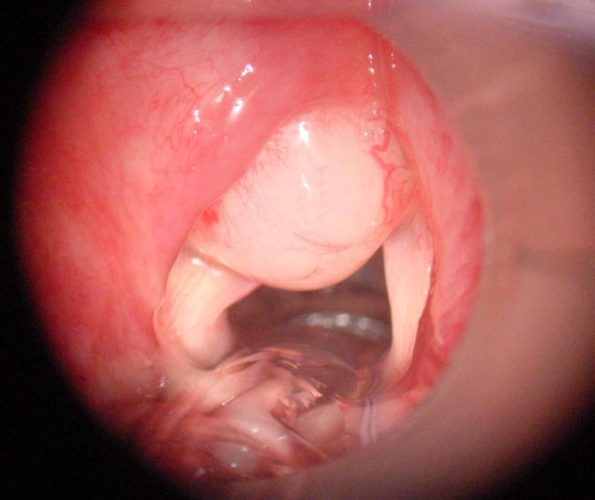
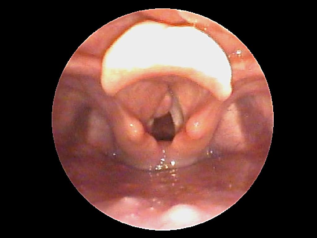
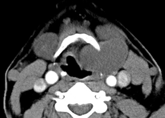
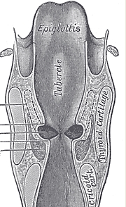
Internal laryngocoeles are limited to the larynx and confined medially by the false vocal cord, and laterally by the lamina of the thyroid cartilage (Figures 1-3).
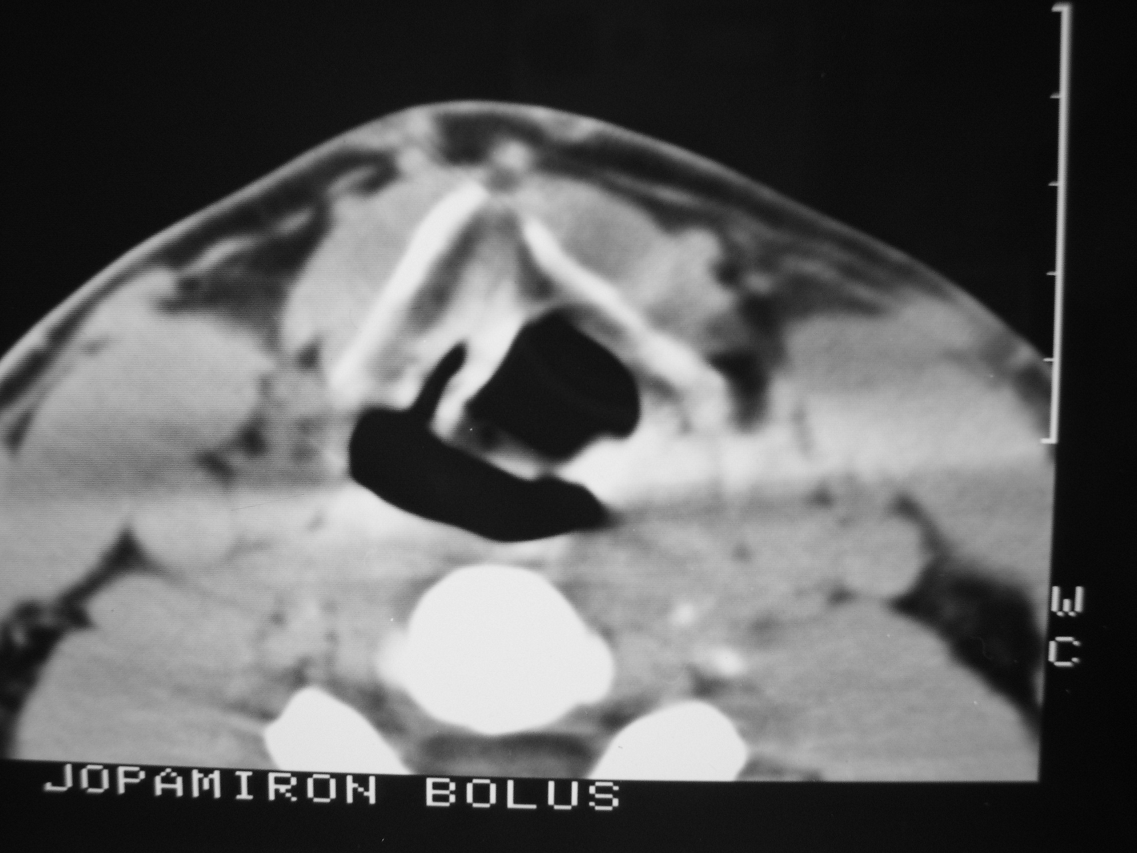
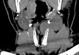
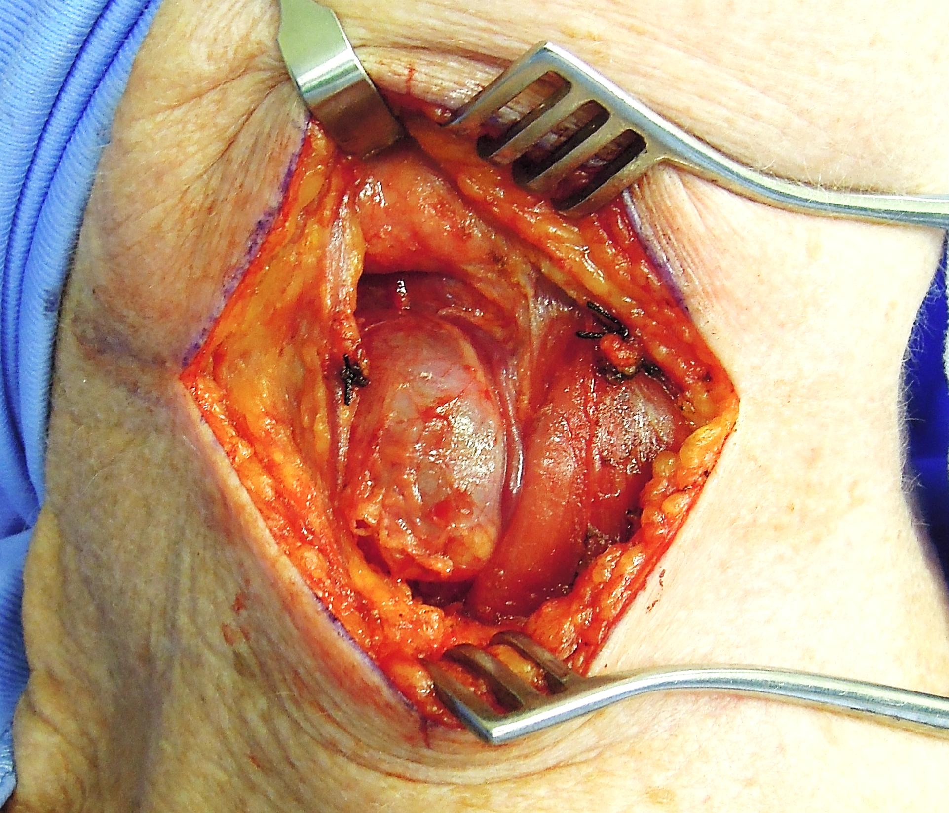
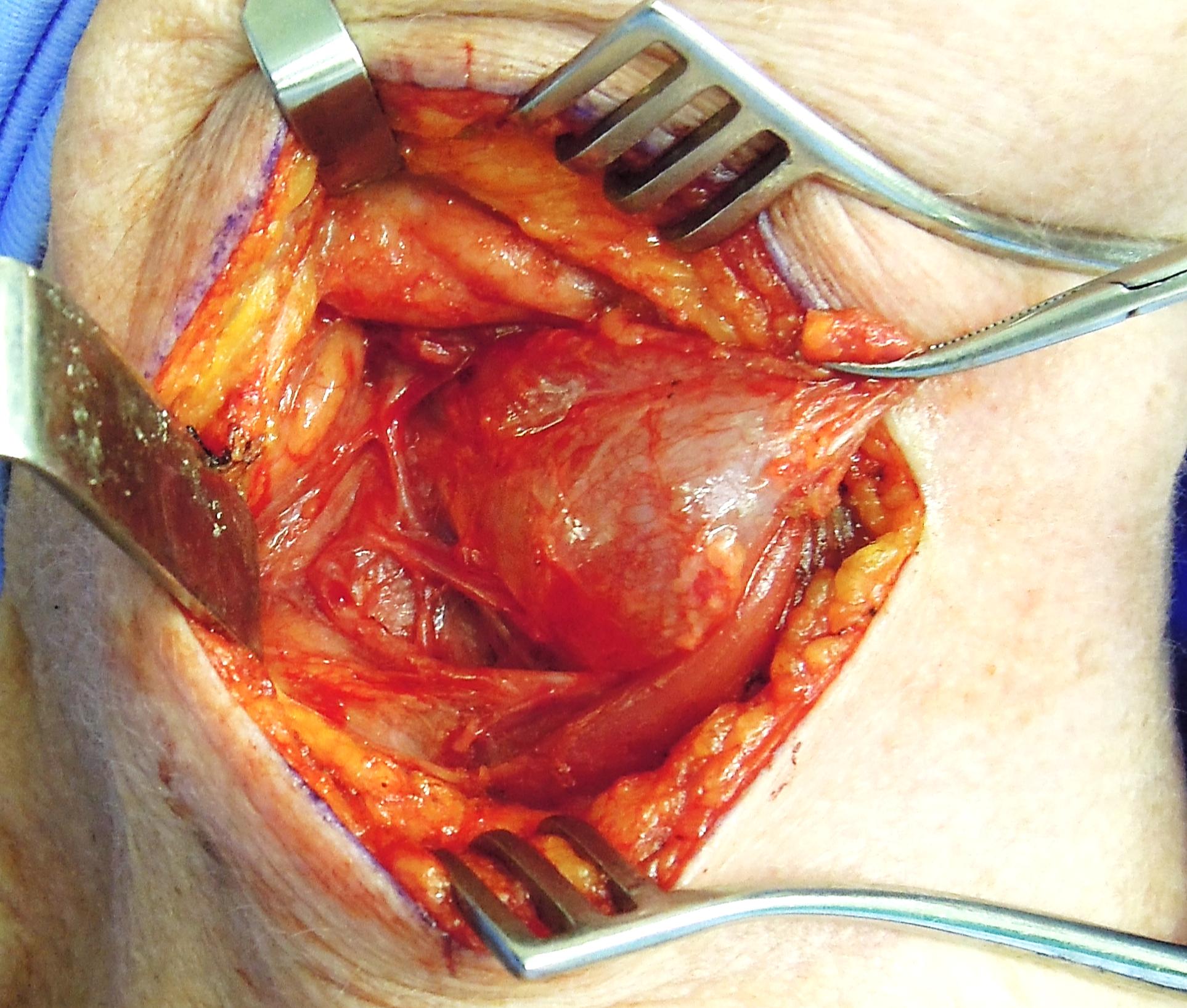
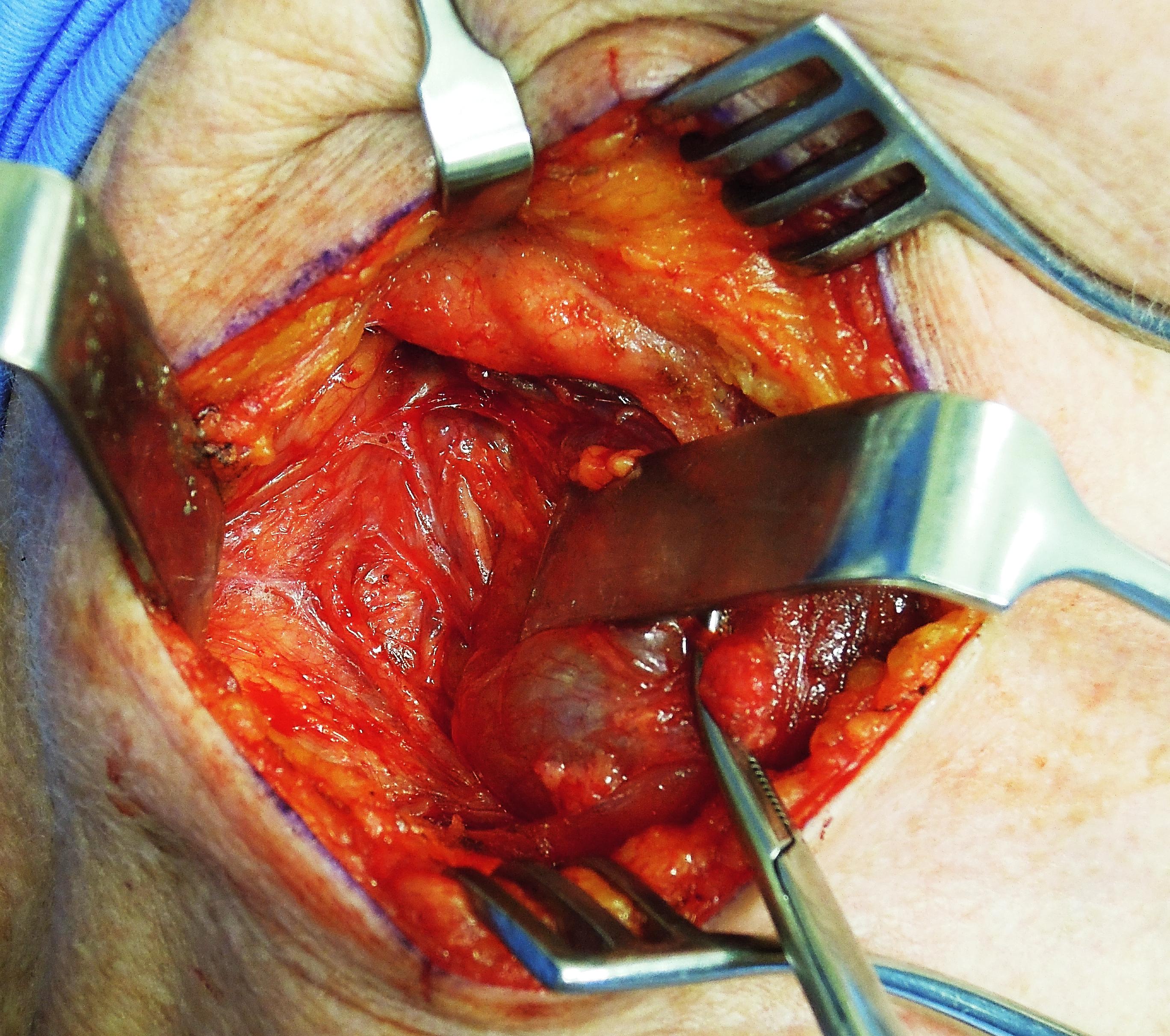
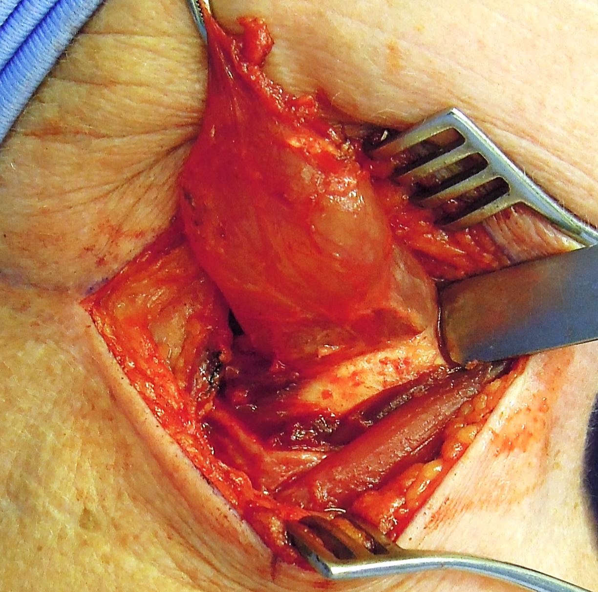
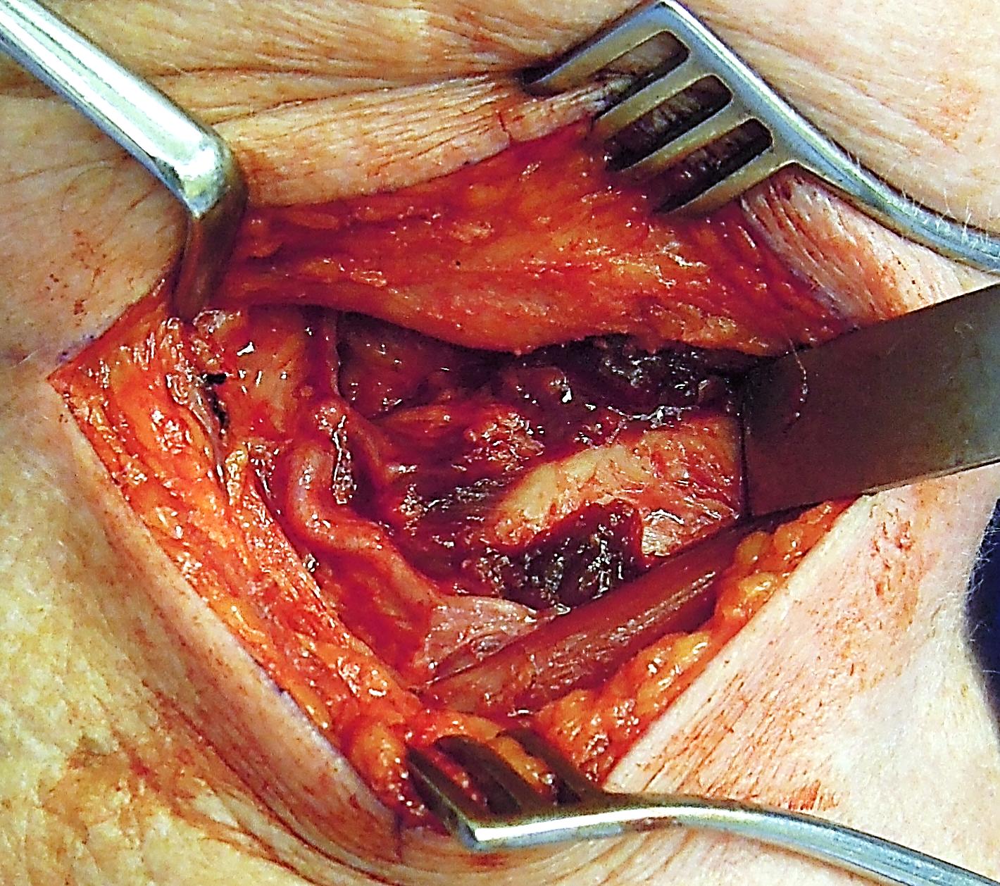
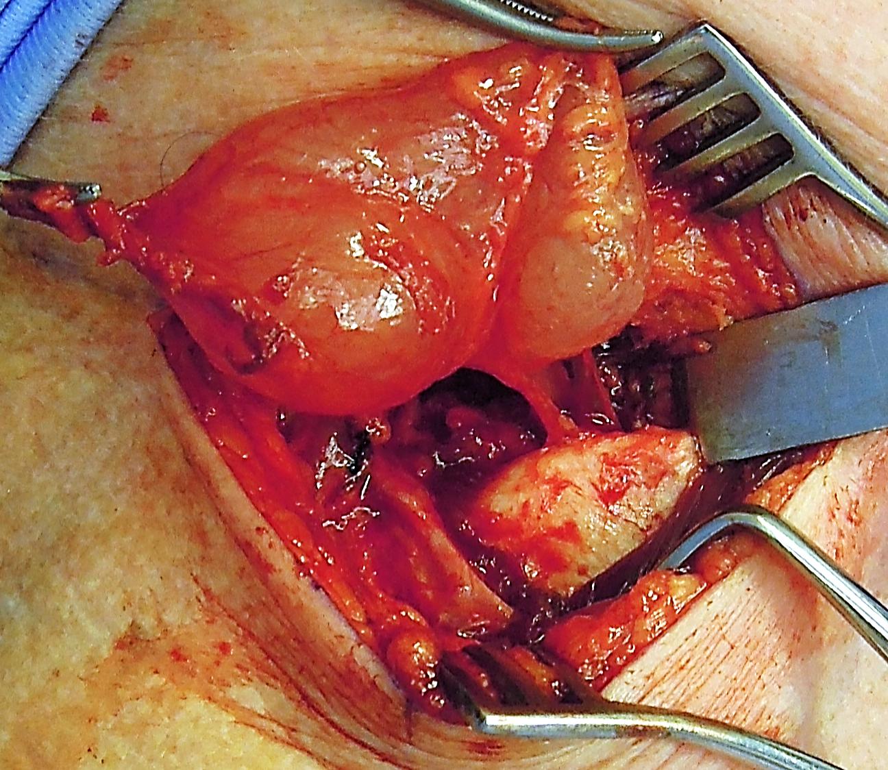
Combined laryngocoeles extend superiorly and laterally into the neck through the thyrohyoid membrane (between the hyoid bone and the superior edge of the thyroid cartilage) in close proximity to the internal branch of the superior laryngeal nerve and superior laryngeal artery (Figures 4, 5, 6).

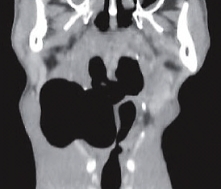
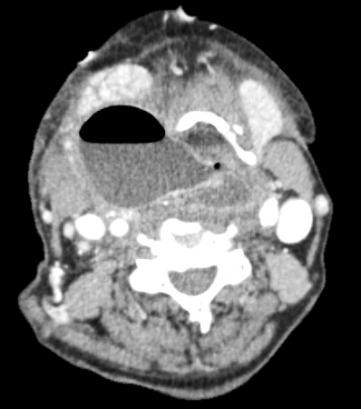
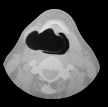
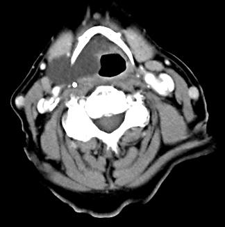
Laryngocoeles are filled with air when they retain a communication with the laryngeal lumen (Figures 1, 4); when they become isolated from the laryngeal lumen they become fluid-filled (Figures 3, 5) or infected (laryngopyocoele) (Figure 6).
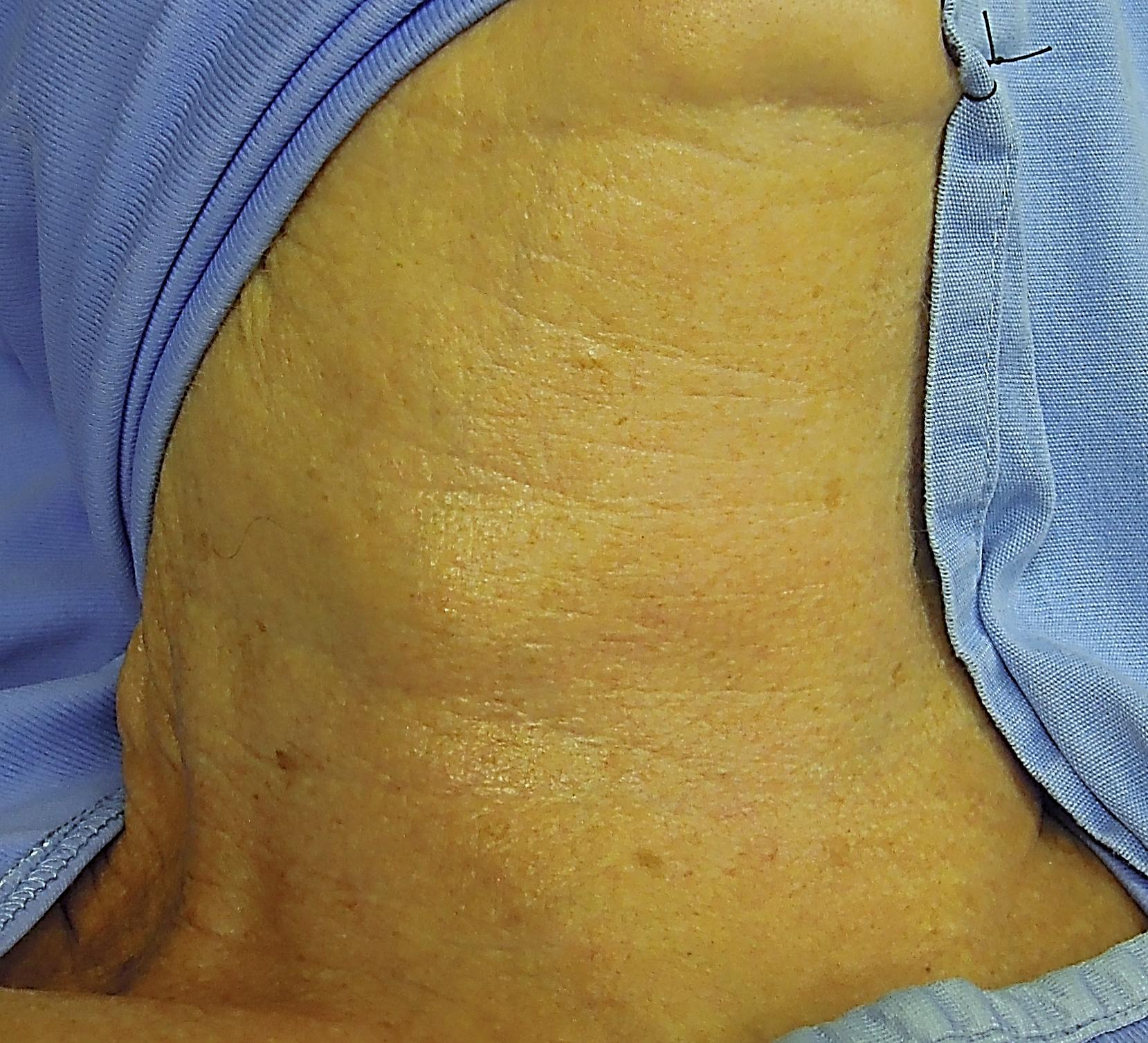
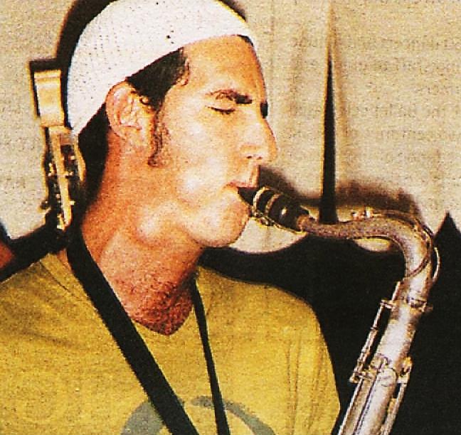
Although not an uncommon incidental postmortem finding, laryngocoeles are generally asymptomatic. Patients may present with voice change or a lateral swelling in the neck overlying the thyrohyoid membrane which may visibly distend when increasing intraluminal pressure e.g. glass blowers and trumpet or reed instrument players (Figures 7a, b).
Patients, especially those with laryngopyocoeles, may present with acute airway obstruction (Figure 6, 8). Occasionally a laryngocoele may be the presenting symptom of laryngeal malignancy obstructing the saccule.
The saccule or appendix of the ventricle is normally present in most larynges. It arises anteriorly in the ventricle and extends superiorly through the paraglottic space with the ventricular fold (false cord) situated medially and the thyroid lamina laterally (Figure 9).
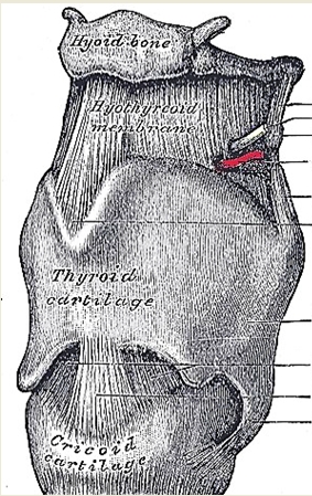
The thyrohyoid membrane extends between the body and greater cornua of the hyoid bone, and the superior rim of the thyroid cartilage. It is pierced by the internal branch of the superior laryngeal nerve and the superior laryngeal branch of the thyroid artery (Figures 10, 11).
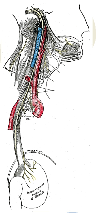
The superior laryngeal nerve is at risk of injury when resecting a laryngocoele due to its intimate relationship to the external component of the cyst. It arises from the ganglion nodosum of the vagus nerve, descends alongside the pharynx, passes behind the internal carotid artery, and divides into external and internal branches. The internal branch crosses the thyrohyoid membrane and pierces it, accompanied by the superior laryngeal artery, and provides sensory innervation to the larynx (Figure 11).
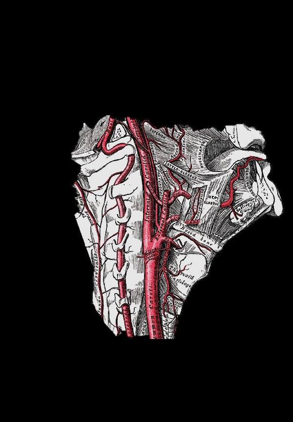
The superior laryngeal artery is encountered during surgery and can either be preserved or sacrificed. It is a branch of the superior thyroid artery (Figure 12).
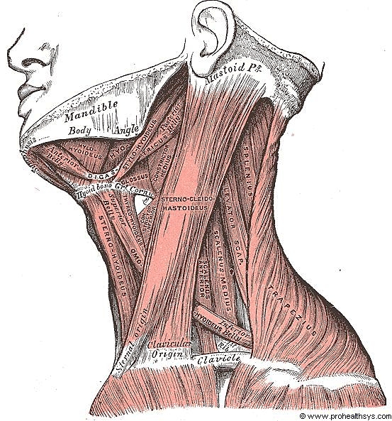
The muscles encountered during resection of the external component of a laryngo-coele are illustrated in Figure 13. The thyrohyoid muscle is draped over the cyst and may have to be divided; the omohyoid can be retracted anteriorly or divided; and the sternomastoid retracted posteriorly.
Imaging
The differential diagnosis of a combined laryngocoele includes a branchial cyst, neck abscess, cold abscess (tuberculosis),
lymphoadenopathy, and a laterally-located thyroglossal duct cyst. An internal laryngocoele can be confused with a carcinoma centered deep in the ventricle which bulges the ventricular fold upwards and medially, and other unusual non-ulcerating masses such as intralaryngeal plasmacytoma, lymphoma and minor salivary gland malignancy.
CT scan will however distinguish between air- and fluid-filled cysts and solid masses. CT evidence of a cyst extending through the thyrohyoid membrane is pathognomonic of a combined laryngocoele. MRI yields similar information.
This depends on the significance of the symptoms and signs, and the size and extent of the laryngocoele. Laryngoscopy is done to exclude the possibility of underlying malignancy in the larynx.
An acutely inflamed combined cyst may first be aspirated percutaneously with a needle and treated with appropriate antibiotics to avoid doing a suboptimal resection in a septic field; needle aspiration may also be employed as an emergency measure to relieve acute airway obstruction.
Internal Laryngocoeles (Figures 1-3)
Small, asymptomatic laryngocoeles do not require surgical intervention. Symptomatic internal laryngocoeles and saccular cysts are widely deroofed/uncapped or excised endoscopically, ideally with CO2 laser. Larger internal laryngocoeles, especially if recurrent, can also be excised by an external approach (see below).

Johan Fagan MBChB, FCORL, MMed
Professor and Chairman
Division of Otolaryngology, University of Cape Town
Cape Town
South Africa
johannes.fagan@uct.ac.za