Total laryngectomy is generally done for advanced cancers of
the larynx and hypopharynx, recurrence following (chemo)radiation, and
occasionally for intractable aspiration and advanced thyroid cancer
invading the larynx.
Although it is an excellent oncologic procedure and secures
good swallowing without aspiration, it has disadvantages such as having
a permanent tracheostomy; that verbal communication is dependent on
oesophageal speech, and/or tracheo-oesophageal fistula speech or an
electrolarynx; hyposmia; and the psychological and financial/
employment implications. Even in the best centers, about 20% of
patients do not acquire useful verbal communication.
Prelaryngectomy decision making
The surgeon needs to consider the following issues before
embarking on a laryngectomy
What will be the tumour resection
lines?As the initial
incisions into the pharynx are done from externally without having the
tumour in view, the surgeon must carefully assess the valleculae, base
of tongue and the pyriform fossae for tumour involvement, so as to
avoid cutting into tumour when entering the pharynx. Involvement of the
base of tongue may also prompt the surgeon to opt for a retrograde
laryngectomy (commencing the laryngectomy at the tracheostomal end of
the specimen). In the absence of CT or MRI imaging, one can palpate and
assess tumour involvement
of the preepiglottic space and base of tongue under general
anaesthesia by placing one index finger in the valleculae, and the
other on the skin of the neck just above the hyoid bone. The fingers
should normally virtually meet, unless there is tumour in the
preepiglottic space or vallecula or base of tongue.
Is thyroidectomy required?
Both hypothyroidism and hypoparathyroidism are common sequelae of total
laryngectomy, particularly following postoperative radiation therapy,
and may be difficult to manage in a developing world setting.
Twenty-five percent of laryngectomy patients become hypothyroid
following hemithyroidectomy; and 75% if post-operative radiation is
added. However both thyroid lobes may be preserved unless Level 6 nodes
need to be resected with subglottic and pyriform fossa carcinoma, or
when there is intraoperative or radiological evidence of direct tumour
extension to involve the thyroid gland.
Will a pectoralis major flap be
required? A capacious pharynx is essential
for good swallowing and fistula speech. Should tumour involve the
hypopharynx, especially when it extends distally towards the
cricopharyngeus, then the expertise has to be available to possibly
augment the pharyngeal repair with a pectoralis major flap. Pectoralis
major muscle flaps are also frequently used to overlay the pharyngeal
repair with salvage laryngectomy to reduce the fistula rate.
Is elective neck dissection required?
With advanced laryngeal squamous cell carcinoma requiring laryngectomy,
elective lateral neck dissection (Levels 2-4), either ipsilateral
(glottic carcinoma) or bilateral (supraglottic, medial wall of pyriform
fossa, bilateral glottic carcinoma) is recommended, with conversion to
modified neck dissection should cervical metastases be found
intraoperatively. Level 6 is included in subglottic and pyriform fossa
carcinoma to clear the paratracheal nodes.
Is the patient suitable for
tracheo-oesophageal speech? This decision is
based on assessment of cognitive function, motivation, financial
ability to pay for replacement speech prostheses, and proximity to
speech services.
Are there synchronous primaries or
distant metastases? Total laryngectomy has
significant morbidity, and should only be done if panendoscopy and
CXR/CT chest exclude metastases or 2nd primaries.
Anaesthesia
Intubation:The operation is done under general anaesthesia. The ENT
surgeon must be present to assist with a possibly difficult intubation.
If a difficult intubation is anticipated, then either do an awake
tracheostomy, or infiltrate skin and trachea with local
anaesthesia/vasoconstrictor, in preparation for a possible emergency
tracheostomy.
Preoperative tracheotomy: Tracheotomy
may have been required for airway obstruction. It is not an independent
indication for postoperative radiation therapy unless tumour was
entered at the time of tracheotomy. If a tracheostomy has already been
done, then ask the anaesthetist to reintubate through the larynx with
an orotracheal tube once the patient has been anaesthetised as this
facilitates dissection in the lower neck and speeds up the surgery.
Perioperative antibiotics:
Commence perioperative antibiotics before putting knife to skin, and
continue for 24 hrs.
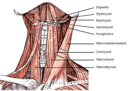
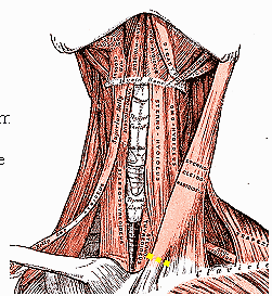
Surgical anatomy
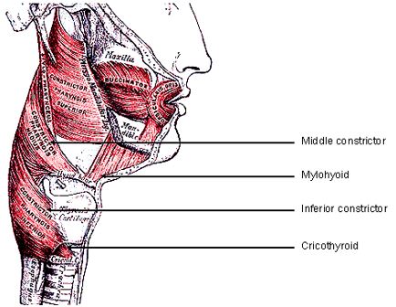
Figures 1 & 2 illustrate all the muscles that will be
divided during laryngectomy.
Figure
1: Supra- and infrahyoid
muscles
Figure
2: Middle and inferior pharyngeal constrictors
Surgical steps
Positioning: Hyperextend the neck
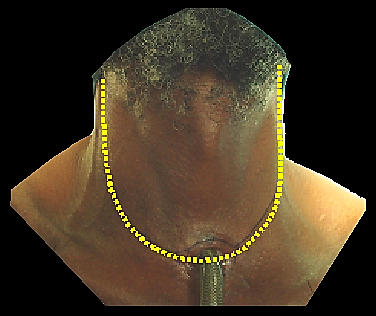
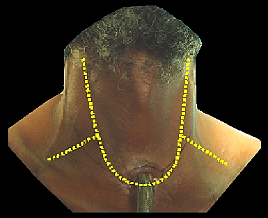
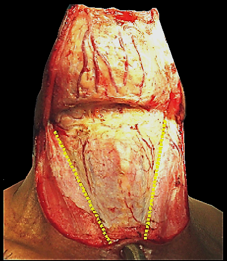
Incisions for apron flap (Figures 3a, b)
The horizontal limb of the flap is placed approximately 2cms
above the sternal notch. An ellipse of skin around a pre-existing
tracheostomy is included with the resection. With a simple laryngectomy
the vertical incision are placed along the anterior borders of
sternocleidomastoid muscles. For a laryngectomy with neck
dissection(s), either a wider flap overlying the sternocleidomastoid
muscles is made (Figure 3a), or a narrow flap
with inferolateral extensions is made (Figure 3b).
The latter has the disadvantage of a trifurcation which is more prone
to wound breakdown and exposure of the major cervical vessels.
Figure
3a: Wide apron flap to
accommodate neck dissectionsFigure
3b: Narrow apron flap for laryngectomy, with
lateral extensions for neck dissections
Flap elevation (Figure 4)
Cut through the superficial layer of investing fascia and
platysma muscles. The platysma is often absent in midline. Take care
not to injure the external and anterior jugular veins
Elevate the apron flap in a subplatysmal plane, remaining
superficial to the external and anterior jugular veins
Dissect the flap superiorly up to approximately 2cms above
the body of the hyoid bone
Figure
4: Elevated apron flap and
incisions through investing layer of cervical along anterior borders of
sternocleido-mastoid muscles
Freeing up the larynx
Free up one side of the larynx at a time. Stand on the side of
neck that is being dissected.
Ligate and transect the anterior jugular veins
suprasternally and above the hyoid
Incise the investing layer of cervical fascia along the
anterior border of the sternocleidomastoid muscle (Figure 4).
Retract the sternocleidomastoid muscle laterally
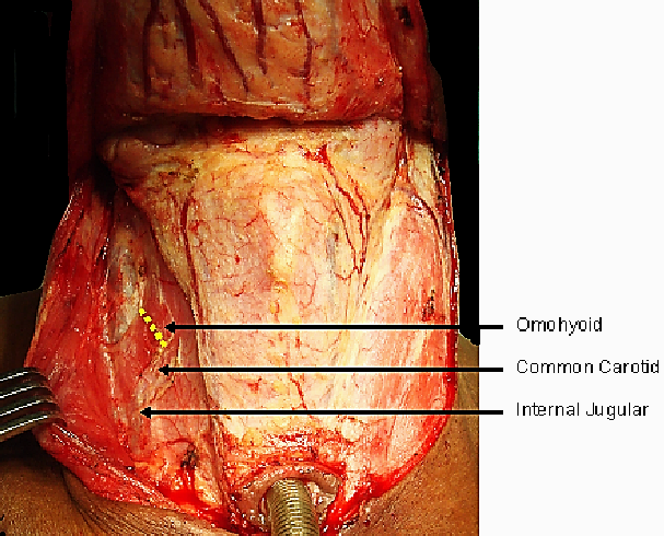
Identify the sternohyoid and omohyoid muscles
Transect the omohyoid muscle medial to where it crosses the
internal jugular vein (Figure 5)
Figure
5: Transect omohyoid along
yellow line
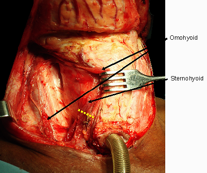
Identify the dissection plane between carotid sheath and
larynx and thyroid gland and open this plane with sharp and blunt
dissection with a finger to expose prevertebral fascia (Figure
6)
Figure
6: Transect sternohyoid muscle
to expose sternothyroid muscle
Transect the sternohyoid muscle with electrocautery
wherever convenient (Figure 6)
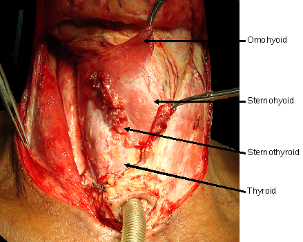
Identify the sternothyroid muscle, and carefully divide it
below larynx (Figure 6). It is a broad, thin
muscle, so take special care not to injure the thyroid gland and its
rich vasculature which is immediately deep to muscle
Carefully elevate and reflect the superior cut end of the
sternothyroid muscle from the thyroid gland using electrocautery
dissection (Figure 7)
Figure
7: Transect & elevate
sternothyroid to expose thyroid glandFigure
8: Divided sternothyroid
retracted to expose thyroid. Line indicates course of dissection of
thyroid gland and along midline of trachea
Divide the thyroid isthmus with electrocautery
Divide and strip the tissues overlying the cervical trachea
anteriorly in the midline to avoid injuring the inferior thyroid veins
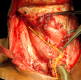
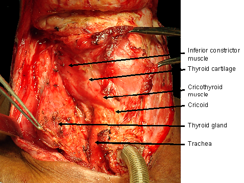
Carefully reflect the thyroid lobe off the trachea, cricoid
and inferior constrictor with electrocautery (Figure 9)
while inspecting for and excluding direct laryngeal tumour extension to
the thyroid gland
Figure
9: Thyroid gland has been
mobilised from larynx and trachea
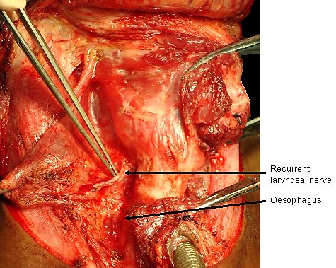
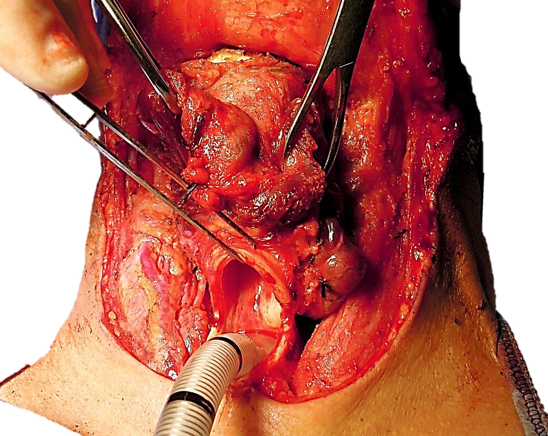
Identify and transect the recurrent laryngeal nerve (Figure
10)
Identify the oesophagus and tracheo-oesophageal groove (Figure
10)
Figure
10: Identify oesophagus, and
divide recurrent laryngeal nerve
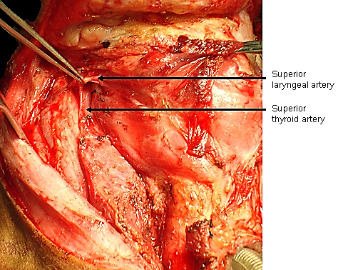
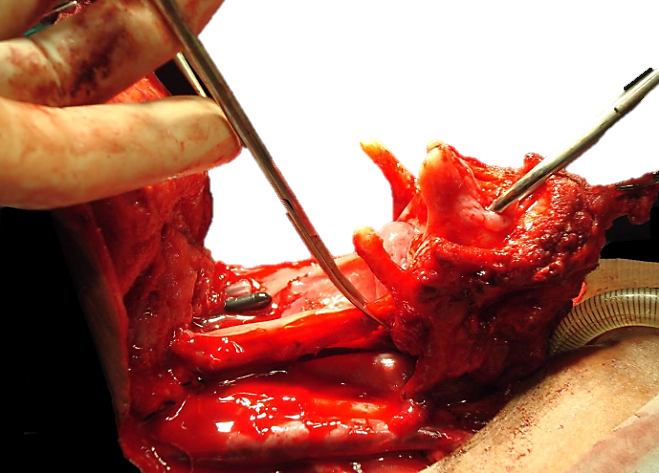
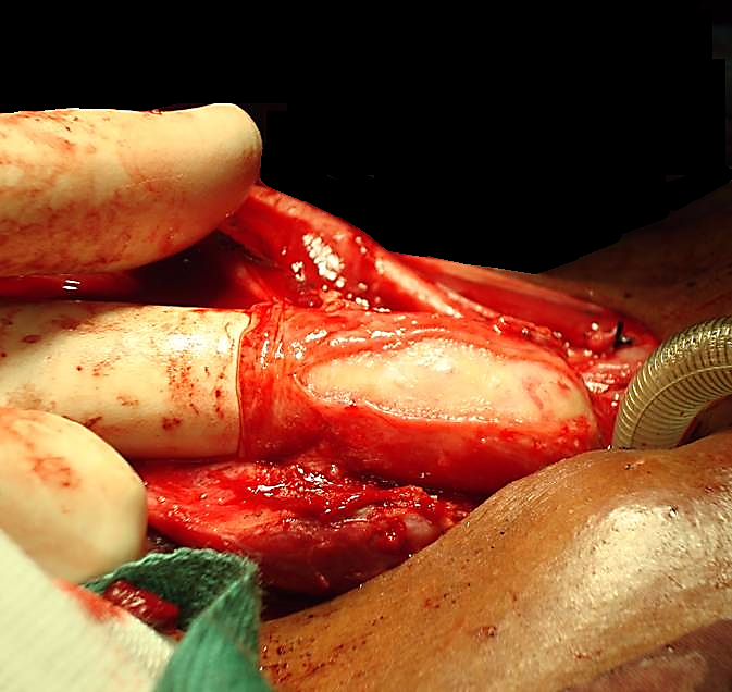
Identify and divide the superior larynx-geal branch of
superior thyroid artery, and reflect and preserve the superior thyroid
pedicle from the larynx (Figure 11)
Identify and divide the superior laryngeal nerve
Figure
11: Identify and divide
superior laryngeal branch of superior thyroid artery
Rotate the larynx to the contralateral side, and identify
the posterior border of the thyroid ala (Figure 12)
Figure
12: Rotate the larynx with a
finger placed behind the thyroid ala
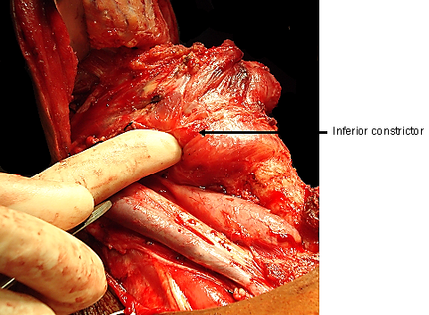
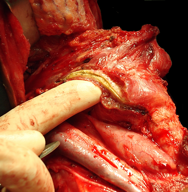
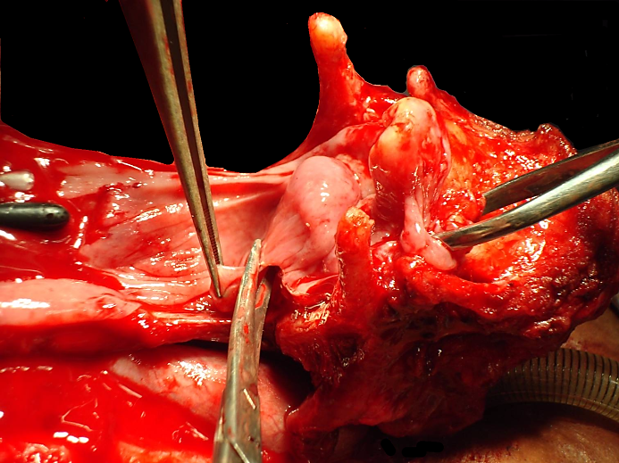
Divide the inferior pharyngeal constrictor muscle and
thyroid perichondrium with electrocautery at, or just anterior to the
posterior border of the thyroid ala (Figure 13)
Figure
13: Divided inferior
pharyngeal constrictor and thyroid perichondrium
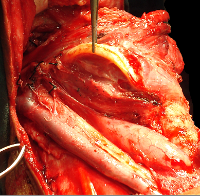
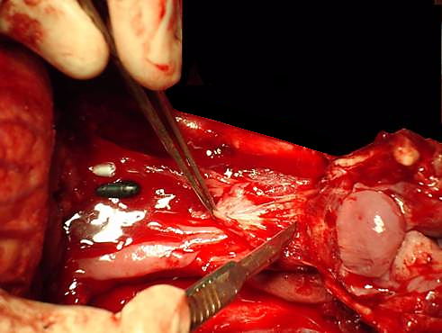
Strip the lateral wall of the pyriform fossa off the medial
aspect of the thyroid ala in a subperichondrial plane with a
swab/sponge held over a fingertip, or with a Freer’s elevator, only
on the side of the larynx opposite to the cancer (Figure 14).
On the side of the cancer, this step is omitted to ensure adequate
resection margins
Figure
14: Pyriform fossa mucosa
stripped from thyroid lamina
The surgeon then crosses to the opposite side of the patient,
and repeats the above operative steps.
Suprahyoid dissection
The following description applies to laryngeal cancer not
involving the preepiglottic space, vallecula or the base of tongue.
When tumour does involve vallecula, pre-epiglottic space and/or base of
tongue, then thepharynx is entered via
the opposite pyriform fossa or a retrograde laryngectomy is done,
commencing the dissection inferiorly at tracheostomy (see later)
Identify the body of the hyoid bone. Remember that the
hypoglossal nerves and lingual arteries lie deep to the greater
cornua/horns of the hyoid bone
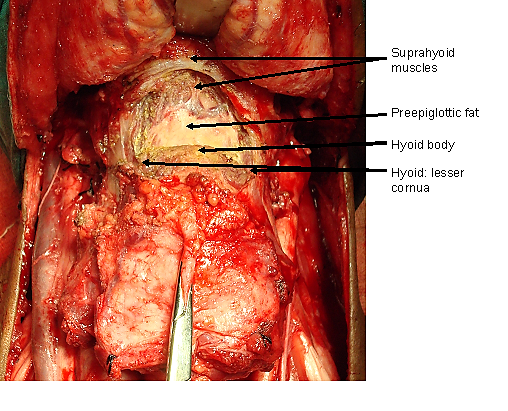
Divide the suprahyoid muscles with electrocautery along the
superior border of the body of the hyoid bone (Figure 15)
Figure
15: Transection of suprahyoid
muscles from hyoid body
Initially do not dissect lateral to the lesser cornua, as
the hypoglossal nerves and the lingual arteries are located deep to the
greater cornua of the hyoid bone
Release the digastric tendon and stylohyoid ligament
and muscle from the lesser cornu of the hyoid. The hyoid then become
more mobile and can be displaced inferiorly, away from the hypoglossal
nerves
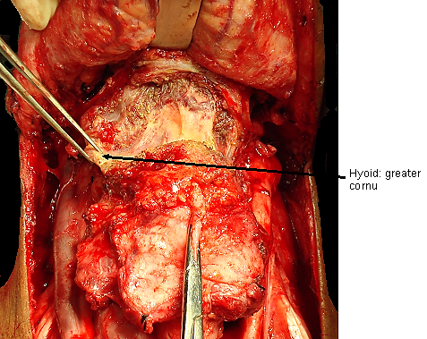
Rotate the hyoid bone to the contra-lateral side, and
identify the position of the greater cornu/horn of the hyoid bone (Figure
16)
Figure
16: Identify greater cornu
The hyoglossus and middle constrictor muscles are next
released from the greater cornu with diathermy
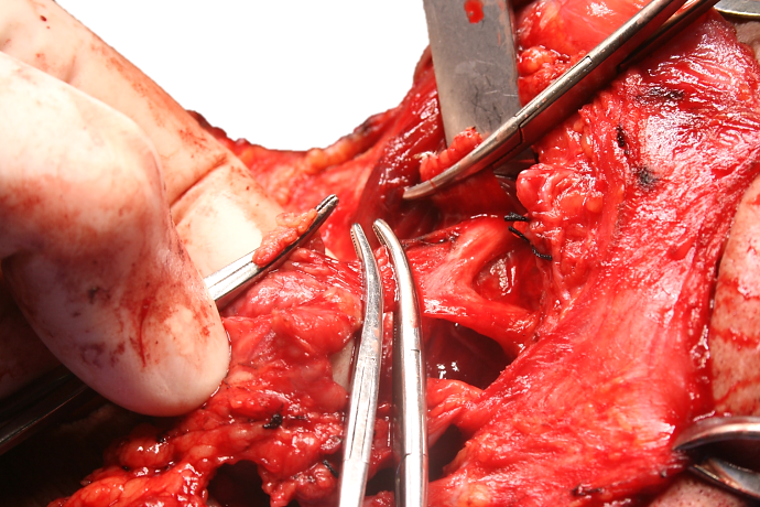
Divide the soft tissue on the medial aspect of the tips of
the greater cornua of the hyoid with scissors to isolate the greater
cornua of the hyoid bilaterally (Figure 17). Hug
the inner aspect of the greater cornua to avoid the hypo-glossal
nerves. If a neck dissection has been done, the hypoglossal nerves will
already be visible
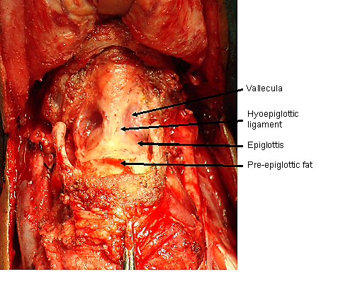
Dissect transversely with diathermy along the superior
margin of the body of the hyoid bone, and along the superior margin of
the preepiglottic space. Identify the hyoepiglottic ligament in the
midline. Dissect along the hyoepiglottic ligament and strip the
vallecula mucosa from the anterior surface of the epiglottis (Figure
18)
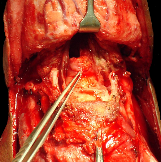
Enter the pharynx by incising the mucosa along the superior
margin of the epiglottis (Figure 19)
A tracheostomy is done at this stage so as to mobilise the
larynx and to facilitate the laryngeal resection
Ask the anaesthetist to preoxygenate the patient
Incise the trachea transversely between the 3rd/4th/5th
tracheal rings or below a preoperative tracheostomy. With a small
trachea, incise the lateral tracheal walls in a superolateral direction
to bevel and enlarge the tracheostoma. Place a few 3-0 vicryl
half-mattress sutures between the anterior wall of the transected
trachea and the skin to approximate mucosa to skin
Puncture and deflate the cuff of the endotracheal tube, and
cut the tube in the pharynx, and remove the distal end of the tube
through the pharyngotomy
Insert a flexible endotracheal tube e.g. armoured tube into
the tracheostoma. Avoid inserting the tube too deeply as the carina is
quite close to the tracheostoma. Fix the tube to the chest wall or
drapes with a temporary suture so that it does not become displaced,
attach the sterile anaesthesia tubing and resume ventilation
Laryngeal resection
Inspect the subglottis through the tracheostoma to ensure
that the tracheal resection margin is adequate
Move to the head of the operating table
Retract the epiglottis and the larynx anteriorly through
the pharyngotomy, and inspect the larynx and the tumour
Commence laryngeal resection contra-lateral to the tumour
using curved scissors with points located anteriorly/upwards so as to
avoid inadvertently resecting too much pharyngeal mucosa
Cut along the lateral border of the epiglottis on the less
involved side, to expose the hypopharynx
Repeat this on the side of tumour, with at least a 1cm
mucosal margin around the tumour
On the less involved side, cut through the lateral wall of
the pyriform fossa and hug the arytenoids and cricoid to preserve
pyriform sinus mucosa (Figure 20). The superior
laryngeal neurovascular pedicle will be transected if not previously
addressed
Repeat on the tumour side
Figure
20: Resect the larynx
preserving maximum amount of pharyngeal mucosa
Join the left and right pyriform incisions by tunnelling
below and cutting the postcricoid mucosa transversely (Figure
21)
Figure
21: Transverse postcricoid cut
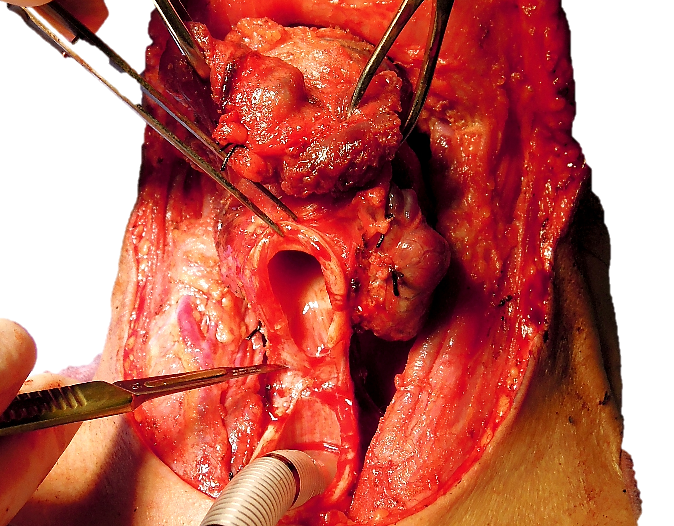
Separate the posterior wall of the larynx (cricoid,
tracheal membrane) from the anterior wall of the oesophagus by
dissecting with a scalpel along the avascular plane between that exists
between oesophagus and trachea/-cricoid (Figure 22).
Take care to stop just short of the tracheostoma.
Figure
22: Dissecting in the
avascular plane between oesophagus and trachea
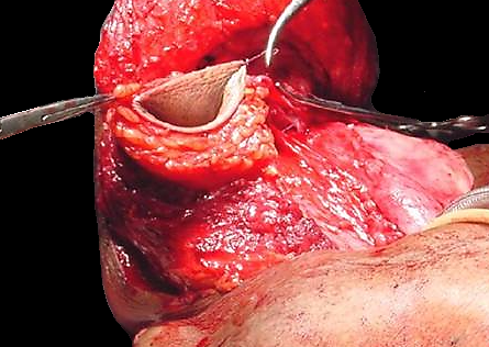
Transect the posterior wall of the trachea, and remove the
larynx (Figure 23)
Inspect the laryngectomy specimen for adequacy of resection
margins, and resect additional tissue if indicated
Figure
23: Transect trachea and
remove larynx
Retrograde laryngectomy
This involves commencing the laryngeal resection inferiorly at
the tracheostomy site; it is recommended when tumour involves the
preepiglottic space and/or base of tongue, in order to ensure an
adequate suprahyoid resection margin. Some surgeons routinely do
retrograde laryngectomy.
Free the hyoid bone and the lateral borders of the thyroid
cartilage as described above
Incise the trachea at about the level of the 3rd/4th
tracheal rings, insert an armored endotracheal tube and remove the
orotracheal tube (Figure 24)
Figure
24: Trachea incised
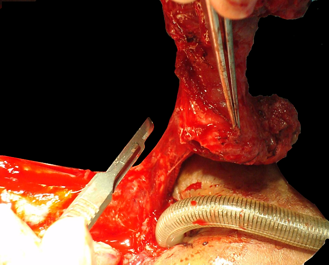
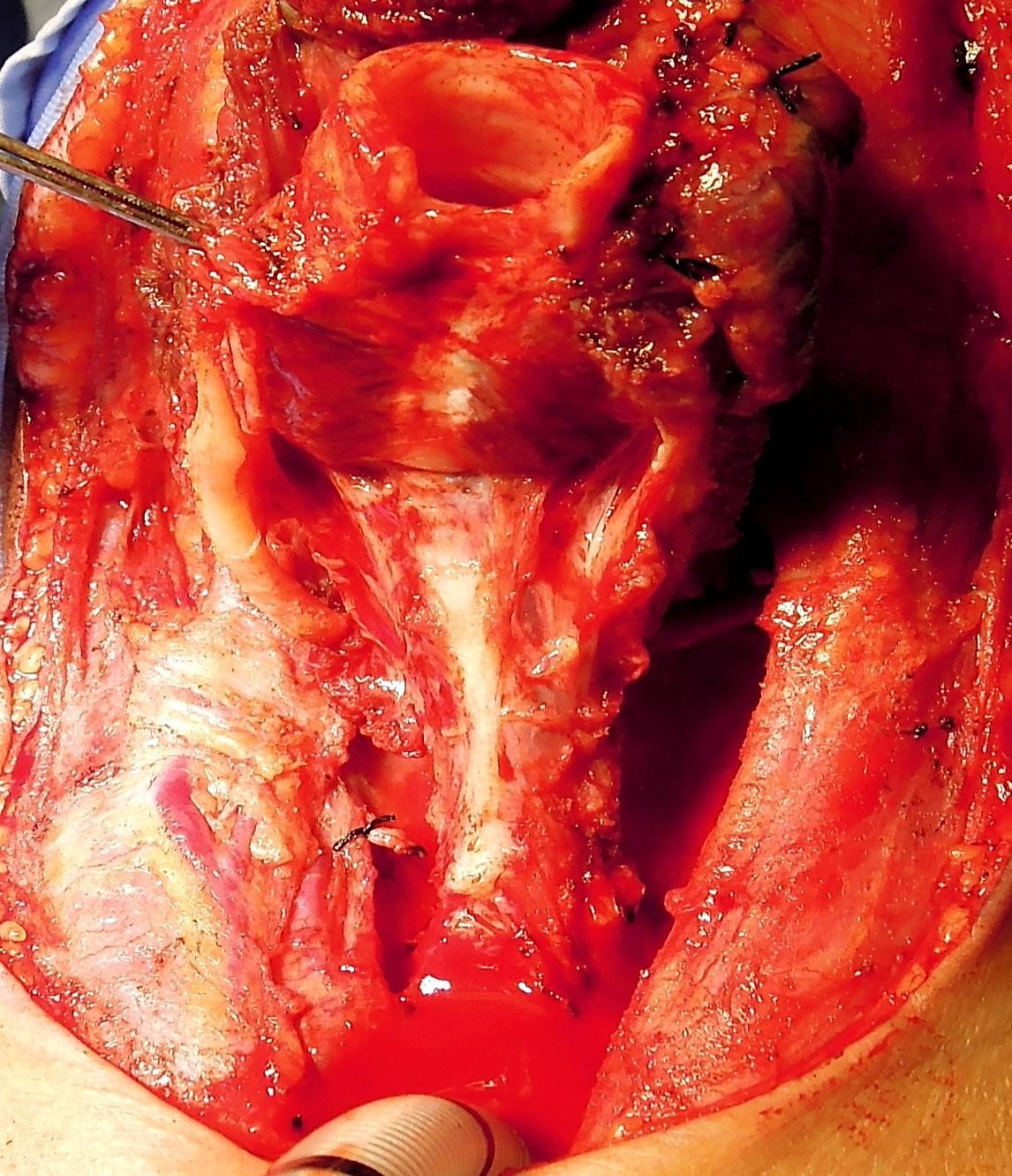
Transect the thin membranous posterior tracheal wall (Figure
25)
Figure
25: Transecting posterior
tracheal wall to expose anterior wall of oesophagus
Find the dissection plane between trachea and oesophagus
and dissect cephalad in this well-defined plane with a scalpel until
the posterior aspect of the cricoid and the posterior cricoarytenoid
muscles come into view (Figure 26)
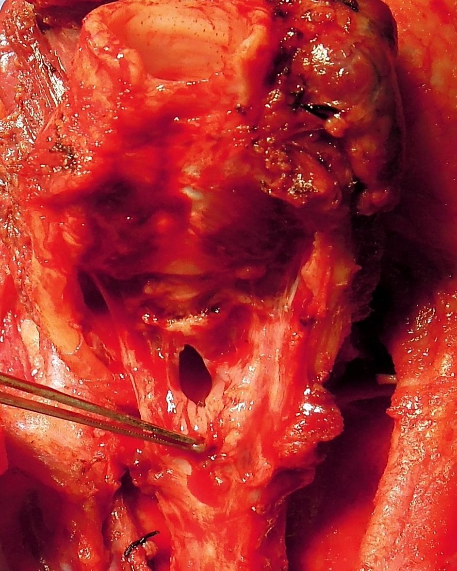
Transversely incise the pharyngeal mucosa about 1cm below
the upper border of the cricoid lamina to enter the postcricoid
hypopharynx (Figure 27)
Extend the incision to the pyriform fossa contralateral to
the cancer
Once the cancer can be seen through the pharyngotomy,
incise the pyriform fossa mucosa on the involved side
By placing an index finger across the vallecula to palpate
the upper extent of the cancer one can proceed to transect
the base of tongue with an adequate margin
Figure
27: Entering the postcricoid
area of the pharynx
Pharyngo-oesophageal myotomy
Optimising speech and swallowing re-quires a capacious and
floppy pharynx
Always perform a pharyngo-oesopha-geal myotomy to prevent
hypertonicity of the pharyngo-oesophageal segment
Insert an index finger into the oesophagus (Figure
28)
Figure
28: Cricopharyngeal myotomy
With a sharp scalpel, divide all the muscle fibres down to
the submucosa, and distally to the level of the trachea-stoma (Figure
28). The myotomy may be done in the midline or to the side
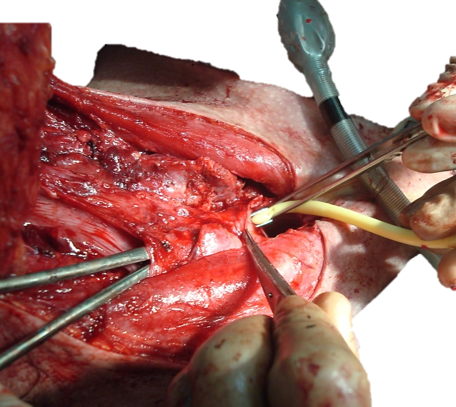
Tracheo-oesophageal fistula
Tracheo-oesophageal speech is the best form of alaryngeal
communication
A tracheo-oesophageal fistula is created before closing the
pharynx
Pass a curved artery forceps through the pharyngeal defect
and along the oesophagus, and tent up the anterior wall of
oesophagus/posterior tracheal wall 5-10mm below the superior margin of
the tracheostoma. Placing the fistula too low makes changing the
prosthesis difficult
Cut down onto the tip of the artery forceps with a scalpel,
and pass the tip of the forceps through the fistula into the tracheal
lumen
Hold the tip of a 14 gauge Foley urinary catheter with the
artery forceps, and pull the catheter through the fistula into the
oesophagus and pass it through the pharyngeal defect (Figure
29). Then advance the catheter down the oesophagus. Avoid
accidental displacement of the catheter by injecting 5ml water into the
bulb and by fixing the catheter to the skin with a suture
Figure
29: Creation of
tracheo-oesophageal fistula
The catheter acts a stent to allow the fistula to mature in
preparation for fitting of a tracheo-oesophageal prosthesis, and is
initially used for stoma-gastric feeding
An alternative method is to insert a speech prosthesis ab
initio, and to feed the patient via a nasogastric tube, or a
catheter passed through the speech prosthesis (Postlaryngectomy
vocal and pulmonary rehabilitation)
Divide the sternal heads of the sternomastoid muscles to
create a flattened peristomal contour and to facilitate digital stomal
occlusion (Figure 30).
Figure
30: Division of sternal heads
of sternomastoid to flatten peristomal area
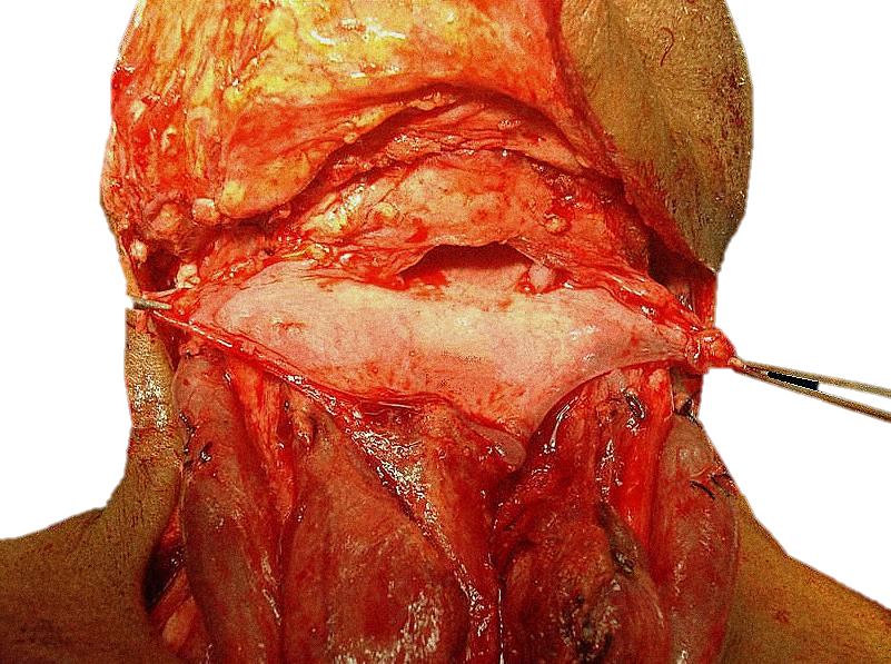
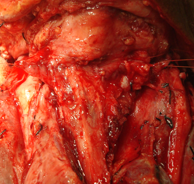
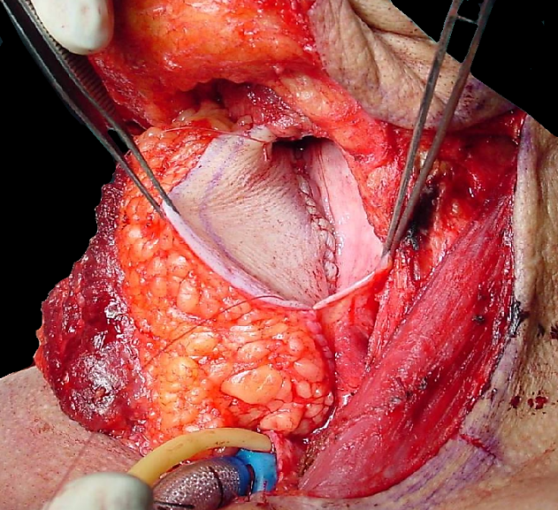
Pharyngeal closure
At least 2.5cm transverse diameter of residual pharyngeal
mucosa is required for primary pharyngeal closure. The teaching that
the minimum pharynx required is that which may be closed over a
nasogastric tube is incorrect, as the neopharynx is then too narrow for
adequate swallowing and voicing
A horizontal/transverse closure is preferred as it
maximises the capacity of the pharynx (Figures 31). Only
if there is undue tension on the suture line, then do T-shaped closure,
keeping the vertical limb as short as possible
Figure
31: Pharynx well suited to a
transverse closure
Take care not to injure the lingual arteries when suturing
the pharynx, as injury to the arteries may lead to necrosis of the
tongue
Figure
32: Completed 1st layer
of transverse closure of pharynx
2nd
layer: 3-0 vicryl running suture of submucosa and muscle
3rd layer: Approximate inferior constrictors and suture
constrictors to suprahyoid muscles with interrupted 3-0 vicryl
Final steps
Ask the anaesthetist to do a Valsalva manoeuvre to detect
bleeding and chyle leaks
If there is excessive, lax suprastomal skin that may
occlude the tracheostomy when the patient flexes the neck, then trim a
crescent of suprastomal skin from the edge of the apron flap
Suture the skin to the edge of the tracheostomy with
half-mattress interrupted 3-0 vicryl sutures
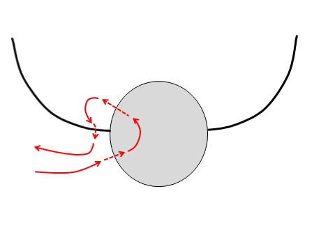
Seal the trifurcation at the lateral edge of the stoma with
a suture as indicated below (Figure 33)
Figure
33: Suture technique to seal
trifurcation between skin and side of tracheostoma
Insert a ¼” suction drain
Irrigate neck with sterile water
Reapproximate the platysma with 3-0 vicryl running sutures
Close the skin with a running nylon suture or with skin
staples
Suction blood from trachea
Insert a cuffed tracheostomy tube, and suture it to skin
Postoperative care
Antibiotics x 24 hours
Omeprazole (20mg/day) via Foley or mouth x 14days to reduce
risk of developing pharyngocutaneous fistulae (unpublished data still in
press)
Chest physiotherapy
Remove suction drains when <50mls drainage per
24hrs (See references)
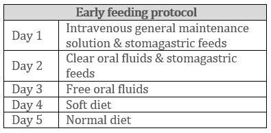
Day 1: Mobilise to chair, remove urinary catheter
Day 2: Commence oral feeding. Early oral feeding is safe,
and does not cause pharyngocutaneous fistulae (See
references)
Day 7: Remove sutures
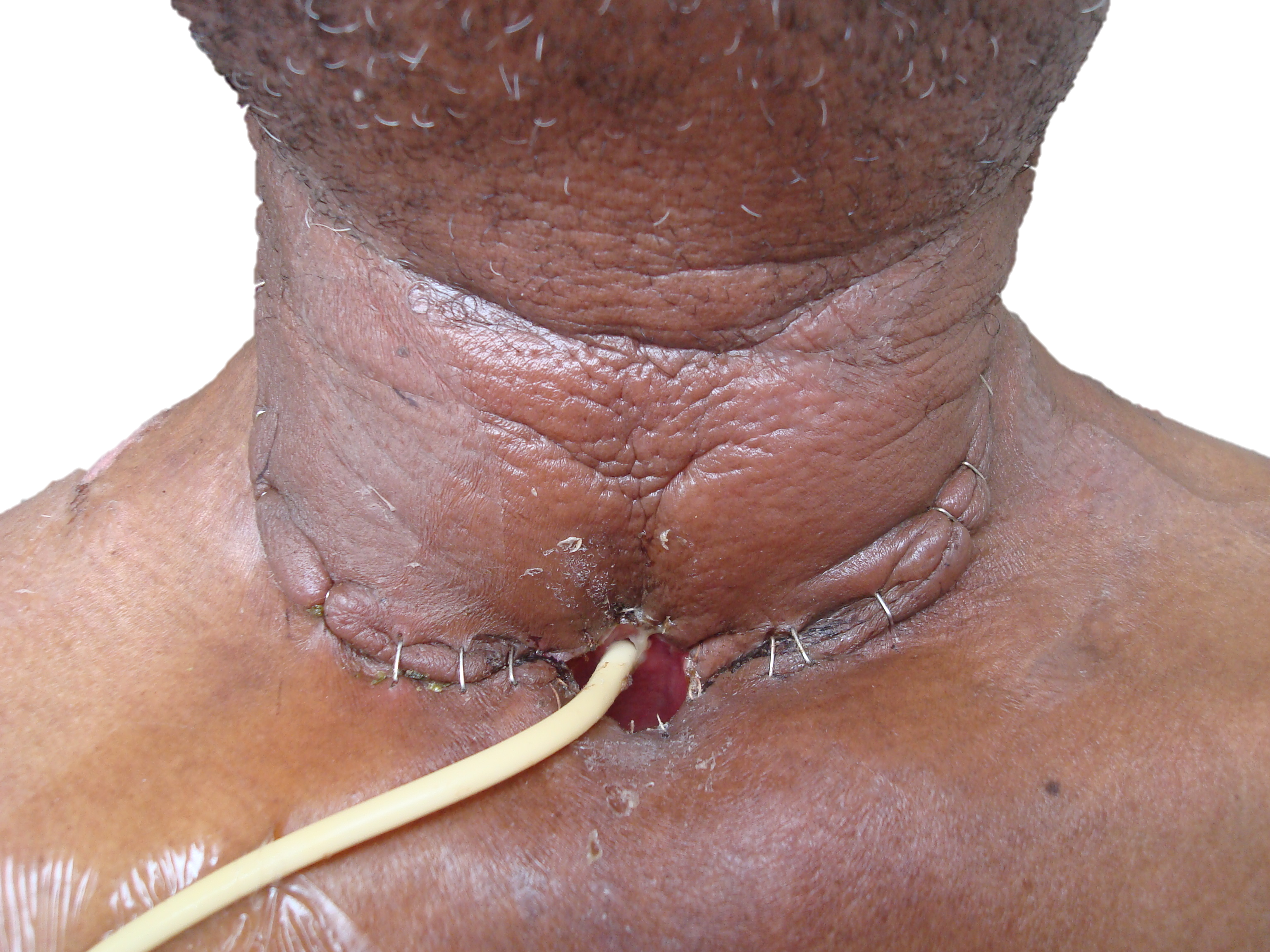
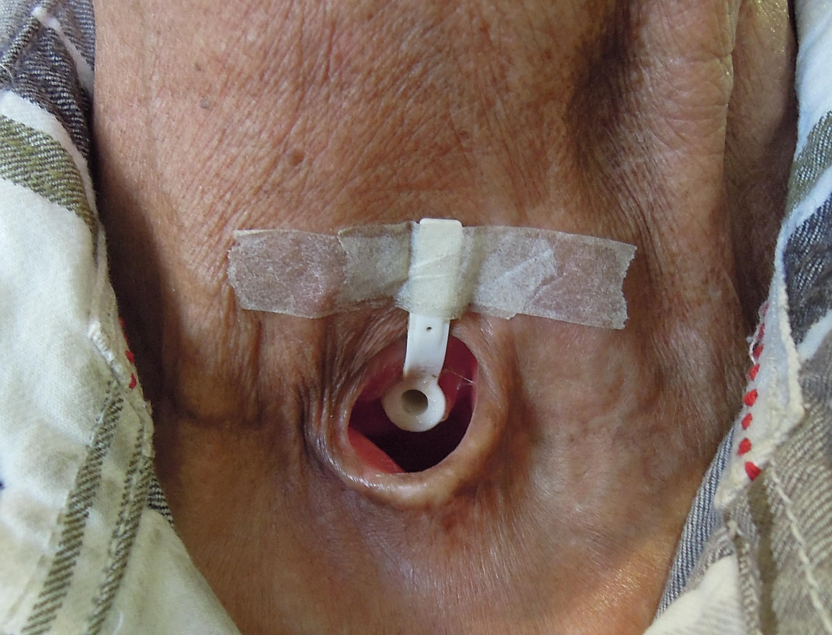
Day 10: Insert speaking valve; no anaesthetic required (Figures
34, 35)
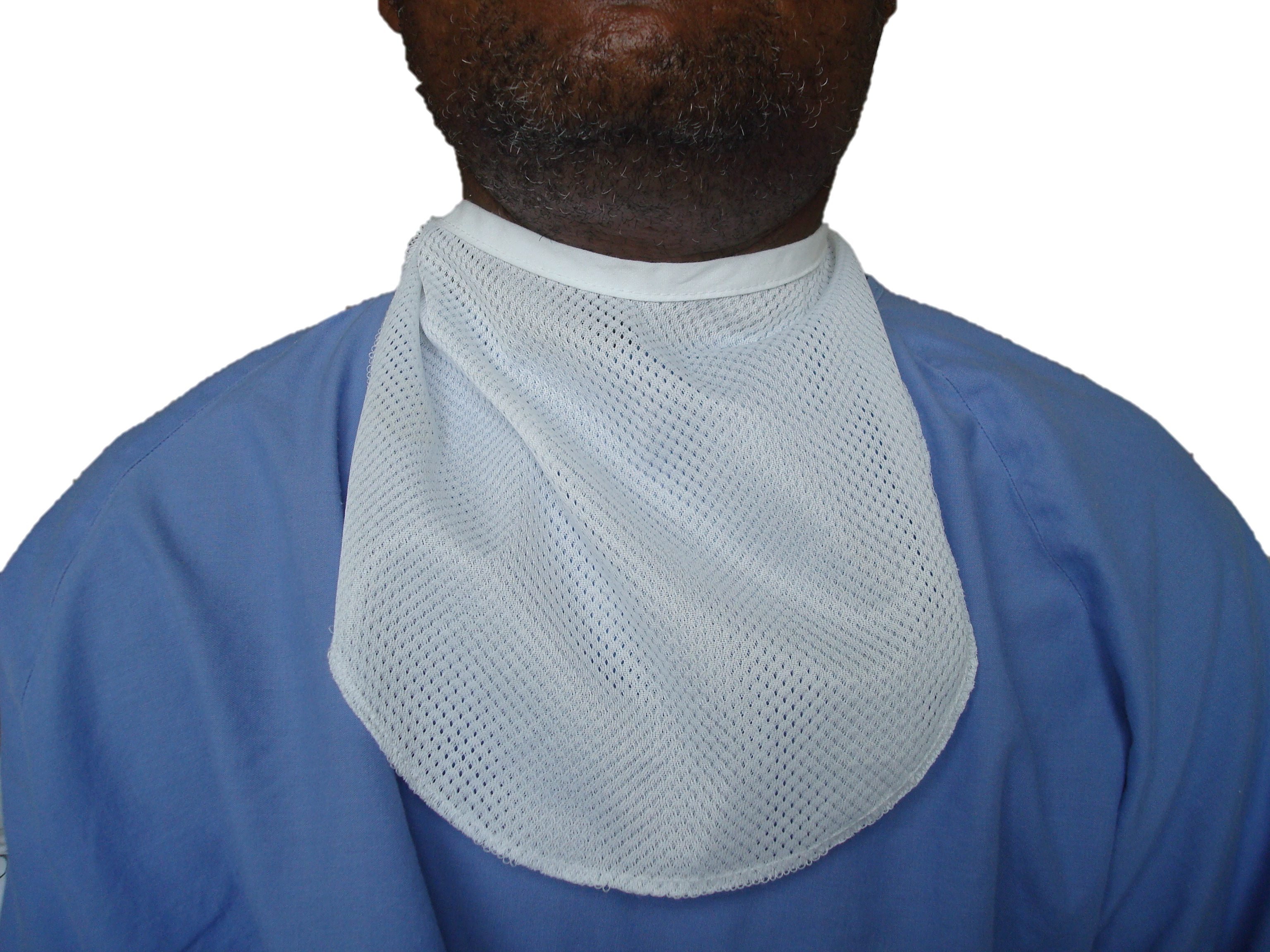
Cover the stoma with a bib (Figure 36)
Figure 34: Stoma and Foley catheter
feeding tube one week following surgeryFigure 35: Speaking valveFigure 36: Bib
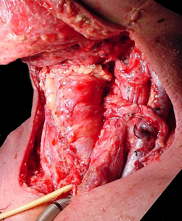
Pharyngeal reconstruction
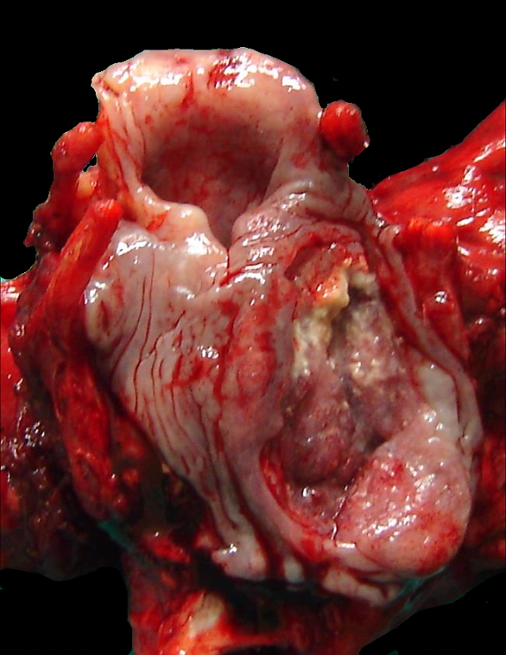
Following resection of large pyriform fossa tumours (Figure
37) or tumours that extend close the cricopharyngeus, or
involve the postcricoid area, only a narrow strip of mucosa may remain
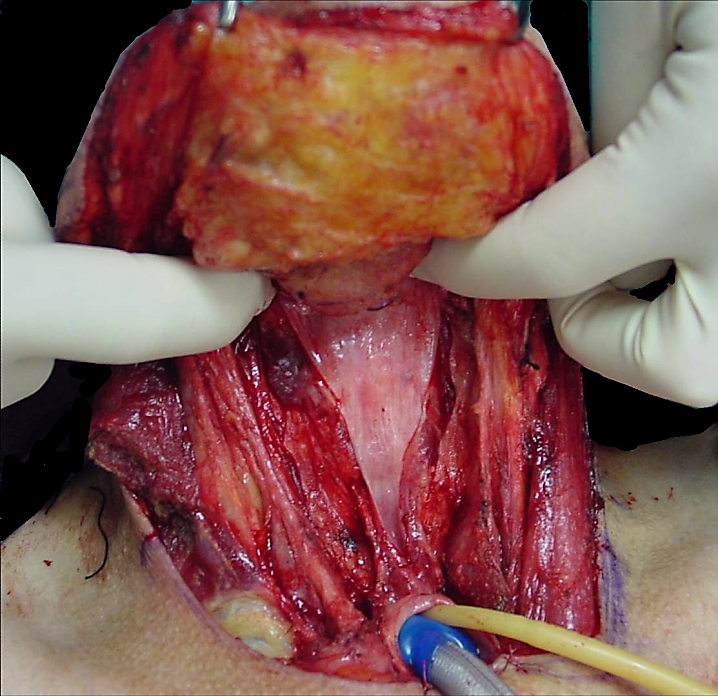
to reconstruct the neopharynx. If the residual pharyngeal mucosal is
<2.5cms in width, then additional tissue is required to avoid
pharyngeal stenosis, dysphagia and poor speech (Figure 38).
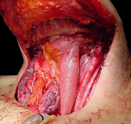
Reconstructive options include pectoralis
major and latissimus dorsi flaps, or microvascular free
tissue transfer flaps (radial
forearm, anterolateral thigh). All these flaps can be used to
augment the pharyngeal repair, or when the pharynx has been completely
resected, may be tubed to entirely replace the pharynx (Figures
39 – 42).
Following pharyngeal reconstruction with a flap, a contrast
swallow X-ray is done on about day 7 to exclude an anastomotic leak
before commencing oral feeding.
Figure 37: Large carcinoma of
hypo-pharynx that will require pharyngeal reconstructionFigure 38:
Insufficient pharyngeal mucosa for primary closure of pharynxFigure 39:
Pectoralis major augmentation of pharynxFigure 40: Tubed pectoralis major flapFigure 41: Tubed
free anterolateral thigh flapFigure 42: Free jejunal flap
Fagan JJ, Lentin R, Oyarzabal MF, S Iaacs, Sellars SL.
Tracheo-oesophageal speech in a Developing World Community. Arch
Otolaryngol 2002, 128(1): 50-53
Fagan JJ, Kaye PV. Management of the thyroid gland with
laryngectomy for cT3 glottic carcinomas. Clin Otolaryngol,
1997; 22: 7-12
Harris T, Doolarkhan Z, Fagan JJ. Timing of removal of neck
drains with head and neck surgery. Ear Nose Throat J. 2011
Apr; 90(4):186-9
Fagan JJ, Lentin R, Quail G. International Practice of
Laryngectomy Rehabilitation Interventions - A Perspective from South
Africa. Curr Opin Otolaryngol Head Neck Surg.
2013 Jun;21(3):199-204
Author & Editor
Johan Fagan MBChB, FCORL, MMed
Professor and Chairman
Division of Otolaryngology
University of Cape Town
Cape Town, South Africa johannes.fagan@uct.ac.za