Forearm injuries
by Pieter Venter & Stephen Roche
Learning objectives
- A basic understanding of the forearm as a single joint.
- Knowledge conerning the clinical anatomy of the forearm.
- An approach to evaluating a patient with a forearm injury clinically.
- An introduction to possible treatment modalities for forearm injuries.
Introduction
The forearm consists of the radius and ulnar bone shafts, linked by the interosseous membrane and spanned between the elbow and wrist joints. The forearm complex functions are to rotate the hand in space (supination/pronation) and the transfer of axial loading forces.
The forearm can be seen as a single joint as structural injuries between the elbow and wrist impact directly on the biomechanics of the forearm. It is essential to screen other areas of the forearm for concomitant injuries in the event of a fracture/dislocation of the radius or ulna
Applied anatomy
- Bones:Radius (radial head proximally), ulna (ulnar head distally)
- Joints: Proximal radioulnar joint (PRUJ), distal radioulnar joint (DRUJ)middle radio-ulnar joint/interosseous membrane (MRUJ)
- Compartments:
- Anterior (flexor): Flexor muscles of hand and fingers, pronators
- Posterior (extensor): Extensor muscles of hand and fingers, supinators (it is important to note that the biceps brachii muscle implants on the proximal radius contribute to supination.
- Nerves: Ulnar nerve (injury will cause claw hand), median nerve (injury will cause Benedict’s sign, radial nerve (posterior interosseous nerve injury (PIN) will cause wrist drop)
- Vessels: Brachial artery divides at the level of the elbow into radial and ulnar arteries.
Clinical findings
History
The patient presents with symptoms of pain, swelling and deformity of the left forearm after a fall on an outstretched hand (FOOSH) from standing height.
Examination
- Look: Evaluate for wounds (abrasions/lacerations), bruises, blisters. Also, look for any deformity/swelling of the wrist and elbow.
- Feel: Neurovascular exam that includes: radial and ulnar pulses, capillary refill time, motor and sensory function of radial, median and ulnar nerves.
Assess for any signs of possible compartment syndrome:.
- Pain out of proportion to the injury.
- Pain on passive stretching of the fingers.
- Gross swelling with tight compartments.
- Move: Loss of normal movement of the forearm due to deformity and pain. Assess hand, wrist and elbow movements for injury.
Additional injuries to note
ALWAYS assess the joint above and below an injury, in this case, the hand, wrist and elbow.
Specific Injuries that occur in the forearm
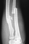
- Monteggia fracture: Proximal ulna fracture with associated radial head dislocation.
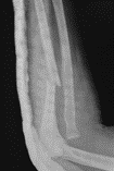
- Galeazzi fracture: Distal ⅓ radial shaft fracture with associated distal radio-ulnar joint (DRUJ) dislocation
Special Investigations Imaging
- ‘2 views and 2 joints’: Always get a minimum of two views (AP and LAT) that include the joint above and below the injury (two joints).
- Describe the X-ray findings:
- Obvious injury or abnormality (fractures/dislocations).
- Fracture pattern (oblique, spiral, transverse and so on): have an impact on stability.
- LARA mnemonic:
- Location (for example, distal ⅓ radial shaft).
- Apposition/displacement (what percentage of the fracture ends are in contact).
- Rotation (for example, are the fragments lying in the same plane?
- Angulation (for example, valgus/varus or dorsal/volar).
 |
 |
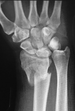 |
| Both bone forearm fracture | Monteggia fracture-dislocation | Galeazzi fracture-dislocation |
Classification
- Descriptive(describing the fracture pattern and whether or not it is an open or closed fracture.
Management
Due to the forearm being a joint, forming an ellipse with the radius articulating over the ulna, the indications for conservative and non-surgical management are limited. Most forearm fractures, therefore, require surgical management.
Non-surgical
- Isolated undisplaced or minimally displaced distal 2/3 ulna fractures (nightstick or defence fractures).
- Moulded below elbow circular plaster of Paris with careful follow up to monitor union and position.
Surgical
- Open reduction and internal fixation(ORIF)
- External fixation
- Intramedullary nailing
Essential takeaways
- The forearm acts as a single joint and injury to one or more structure impacts significantly on its biomechanics.
- Always assess the hand, wrist and elbow for associated injuries
- Do a proper neurovascular examination and note specific clinical findings – even if they are normal.
References
Dumontier C, Soubeyrand M. The Forearm Joint. In: Bentley G (eds) European Instructional Lectures. European Instructional Lectures, Vol 13. Springer, Berlin, Heidelberg https://link.springer.com/chapter/10.1007%2F978-3-642-36149-4_14#citeas. Published online 16 February 2013. Accessed 22 October 2019.
LaStayo C, Lee M. The forearm complex: anatomy, biomechanics and clinical considerations. Journal of Hand Therapy. https://www.sciencedirect.com/science/article/pii/S0894113006000421?via=ihub. Published 22 November 2006. Accessed 22 October 2019.
Moore D. Galeazzi Fractures. Orthobullets. https://www.orthobullets.com/trauma/1029/galeazzi-fractures. Accessed 22 October 2019.
Rosenwasser M, Hess A, Mighell M, Elsaied M, Morales J. Monteggia Fractures. Orthobullets. https://www.orthobullets.com/trauma/1024/monteggia-fractures. Accessed 22 October 2019.
Strauch R, Steimle J, Yip, Dalla-Rosa J, Orthobullets Team. Radius and Ulnar Shaft Fractures. Orthobullets. https://www.orthobullets.com/trauma/1025/radius-and-ulnar-shaft-fractures. Accessed 22 October 2019.
Assessment
A 24-year-old male patient presents with a left sided Monteggia fracture-dislocation after a fall from a height. He complains of pain in his forearm and elbow. Your examination shows that he has a drop wrist (cannot dorsiflex his wrist). Which nerve is most likely to be injured in this fracture pattern?
- Ulnar nerve
- Posterior interosseous nerve
- Median nerve
- Anterior interosseous nerve
- Lateral antebrachial cutaneous nerve
- Incorrect. The radial nerve divides into two branches in the antecubital fossa (deep/motor and superficial/sensory) with the deep motor branch passing through the heads of the supinator muscles to become the posterior interosseous nerve (PIN) which winds around the radial neck and innervates the muscles of the extensor (posterior)compartment of the forearm, which are responsible for wrist extension.
- Correct. The radial nerve divides into two branches in the antecubital fossa (deep/motor and superficial/sensory) with the deep motor branch passing through the heads of the supinator muscles to become the posterior interosseous nerve (PIN). It winds around the radial neck and innervates the muscles of the extensor (posterior) compartment of the forearm, which are responsible for wrist extension.
- Incorrect. The median nerve enters the forearm anterior to the lateral humeral epicondyle to supply the volar (flexor) compartment of the forearm.
- Incorrect. The anterior interosseous nerve (AIN) is a terminal branch of the median nerve.
- Incorrect.The lateral antebrachial cutaneous nerve(LABC) is a purely sensory nerve that originates from the musculocutaneous nerve and does not have any motor function.