Proximal upper limb fractures in children
by Anria Horn
Learning Objectives
- Identify and describe common upper limb fractures in children.
- Understand how to manage these fractures conservatively.
Clinical assessment
Children tend to sustain fractures in predictable locations. The most common mechanism of injury for all upper limb fractures is a fall on an outstretched hand (FOOSH). The most common fractures are clavicular > distal radius (+/- ulna) > supracondylar fractures. The growth plate is a weak spot in children’s bones, and fractures often occur through and around them. It is important to know what normal growth plates and ossification centres look like to identify these fractures.
Clavicular fractures
Clavicular fractures are the most common in children and are a common birth injury. One should exclude neurovascular injury as there is close proximity to the brachial plexus and large vessels.
Management
Conservative management, involving a collar and cuff or arm sling, is indicated for all clavicular fractures in children. However, conservative management should not be applied to open clavicular fractures or those associated with vascular injury (rare).
Humeral fractures
Spiral humerus fractures in small children are suspicious for non-accidental injury. In all of these fractures, one should exclude axillary nerve (proximal humerus) or radial nerve (midshaft) injury.
Management
- These fractures can mostly be treated with simple immobilisation in a collar and cuff or U-slab. Three weeks of immobilisation is usually adequate.
- A large degree of angulation can be accepted. Remodelling is robust and, as a non-weight bearing limb, small amounts of residual deformity are acceptable
Fractures around the elbow
There are many ossification centres around the elbow appearing at different timesas the child matures. These ossification centres may look like fractures to the inexperienced eye. The acronym CRITOE/ CRMTOL is useful to remember the ossification centres and when they appear. The appearance of the ossification centres is summarised in the table and images below.
| C | Capitellum | 1–2 years |
| R | Radial head | 3–4 years |
| I/M | Internal/medial epicondyle | 5–6 years |
| T | Trochlea | 7–8 years |
| O | Olecranon | 9–10 years |
| E/L | External/lateral epicondyle | 11–12 years |
Management principle: It is important to note that you should never use a circular cast for elbow fractures, always a backslab.
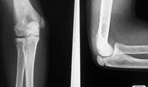
Supracondylar fractures
There is a weak spot above the humeral condyles at the level of the olecranon and coronoid fossae. Injuries here are typically FOOSH.
Clinical examination and investigations
- Diffusely swollen elbow
- Exclude vascular and neurological injury
- Arteries: Brachial artery. Check radial pulse and hand perfusion.
- Nerves: Median > ulnar > radial.
- X-rays: AP and lateral and a contralateral views of the elbow or both in cases of uncertainty. The Gartland Classification (Grade I–III) is used to grade these fractures.
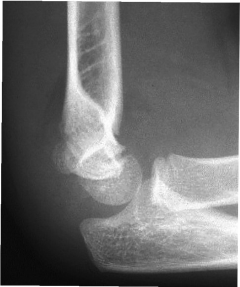
I. Undisplaced fracture
Here one may only see the fat pad sign. The fat pad sign is appreciated on the lateral view X-ray. Usually, an anterior fat pad can be seen. The presence of a posterior fat pad on X-ray is abnormal.
II. Partially displaced with intact posterior periosteal hinge
If the anterior humeral line crosses the capitellum, no reduction is required, and management is as for Type 1.
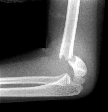
III. Completely displaced fracture
Here there is a completely displaced fracture (See below: the anterior humeral line does not pass through the capitellum). There is a high risk of neurovascular injury (an orthopaedic emergency). These patients require urgent reduction and referral for operative management.
Complications may include:
- Compartment syndrome of the forearm. Also known as Volkmann’s ischaemia.
- Neurological injury (the median nerve most commonly injured)
- Malunion leading to gunstock deformity (cubitus varus)