Hand infections
by Ian Koller, Pieter Jordaan & Neil Kruger
Learning objectives
- Early recognition and appropriate action save hand function.
- Severe hand infections need STAT empiric IV antibiotics PRIOR to theatre and PROMPT referral for surgical drainage.
- All patients with diabetes and HIV should be given Augmentin as their antibiotic treatment.
- Oral flucloxacillin is appropriate for local infections not requiring IV antibiotics.
- Antibiotics cannot penetrate abscesses – these need to be drained surgically.
- Hand sepsis in a diabetic or immunocompromised is an emergency.
- Specimens, preferably tissue, should be sent for microscopy and culture with every infection requiring surgical management.
- Hand infections cause stiffness, which must be proactively managed to restore function.
Introduction
Definition
Hand infections cover any bacterial, viral, mycotic or other agent infecting the hand. There are 5 main hand infections, all of which have a predominant bacterial (staph or strep) aetiology.
| Main hand infections |
|---|
|
The first two are severe hand infections that require immediate empiric IV antibiotics (covering staph and strep – e.g. Cloxacillin), admission to hospital and theatre booking on the emergency list for incision and drainage.
Background
Hand infections are very common in the general population, especially in professions involving work where cuts and scrapes on the hands are common (e.g. manual labourers, such as brick layers), as these are a portal for bacterial entry.
Infective tenosynovitis of flexor tendons
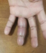
This is less common than other hand infections and presents with deep seated pain along the length of the volar aspect of the finger extending up to the distal palm. It is acutely painful and the finger is kept in a position of mild flexion. Patients will resist passive extension of the finger, as any movement of the tendon within the sheath is severely painful.
The infection can spread proximal to the carpal tunnel and can then spread along other tendons. It is very important to examine the whole hand and forearm to exclude proximal spread of infection.
| Kanavel’s 4 signs of flexor tenosynovitis |
|---|
|
Four classic signs described by Kanavel to aid the diagnosis of an infective tenosynovitis.
Management
All patients with this diagnosis need immediate IV antibiotics (empirically before theatre!), admission to the ward and booking on the emergency list for incision and drainage.
Surgical technique
A 1.5cm–2cm transverse incision just proximal to the A1 pulley an oblique incision of similar length is made midway over the volar aspect of the distal phalanx to provide access to the proximal and distal aspects of the flexor tendon sheath. Proximally a small feeding tube (NGT or drip cannula) is introduced into the sheath and copious amounts of water or saline are syringed through the sheath (must see it run out of the distal incision continuously), until the solution is completely clear and the sheath rid of all pus/murky/turbid fluid or collections. If proximal spread has occurred, this should also be dealt with surgically.
Deep palmar space infection
An infection of the deep palmar space where the pus gets trapped deep in the hand is an emergency. Penetrating injury to the palm is a common cause. Presentation and recognition of this is often delayed as it is somewhat concealed and the obvious fluctuance not there. The hand appears very swollen, but the swelling may appear to be located dorsally, rather than palmar. The swelling may be more radial or ulnar, as this space is separated by a fibrous sheath running longitudinally along the 3rd metacarpal. The natural concavity of the palm may be lost as the pus collects between the volar flexor tendons and the metacarpals dorsally.
Management
Admission to hospital, immediate empiric IV antibiotics, and booking on the emergency list for incision and drainage is required. Incisions are over the point of maximum fluctuance, or over the point of maximum swelling, trying to stay longitudinal or oblique. Use a scalpel through the skin and then a blunt dissecting scissors to access the abscess cavity (less risk to nerves). Perform a thorough debridement of any devitalised tissue and copiously irrigate the cavity to clear of all the pus and debris. Apply a Betadine-soaked dressing into the cavity and wrap it well with velband and kling bandage. Prescribe pain medication (Paracetamol, ibuprofen and tramadol) and antibiotics (Cloxacillin/Co amoxiclav 1g 6hrly) until MCS results are back. Do a wound inspection at 3 days after the initial surgery to assess response and need for re-debridement.
Deep palmar space infection
This presents as a swelling on the volar aspect of the distal palm between the fingers, classically causing them to splay apart due to the space occupying effect. It may break through dorsally onto the extensor surface, where it often may blister and spread along the looser dorsal fascial planes.
Management
This may be managed as day case surgery with discharge home the same day if no systemic signs of sepsis are present. Management consists of a longitudinal volar incision between the fingers from the base of the webspace, big enough to admit a finger to deloculate and release the pus. Avoid crossing the apex of the webspace, as this can cause scarring and contracture of the fingers, decreasing their abduction ability or span. Dress the wound as for other abscesses. Prescribe pain and anti- inflammatory medication and flucloxacillin 500mg 6hrly for 1 week or until cleared. Do a wound inspection at 3 days after the initial surgery to assess response and need for re-debridement.
Felon
A felon is a pulp space abscess of the distal phalanx of any finger. It is often secondary to small cuts or a penetrating injury to the finger. The patient usually presents with a swollen terminal digit and acute, severe throbbing pain which often keeps them awake at night. Unless released, this pain will worsen due to ischaemia and then suddenly abate as the tissues necrose. The fingertip contains numerous enclosed small fibrous compartments which cannot expand much and hence rapidly become painful with the swelling. The key to successful management lies in early recognition and surgical release thereof. If unchecked, the infection may cause necrosis of the tissues and osteomyelitis of the terminal phalanx. Always do an X-ray to check beforehand as once osteomyelitis has set in it is very difficult to save the phalanx and ablation should be considered.
Management
If no osteomyelitis on X-ray, a longitudinal incision over the point of maximal fluctuance should be made under digital block. It is important to release the fibrous septae to decompress the affected area. Remainder of management is as for a webspace abscess above. Do not excise a diamond as this results in significant scarring, takes longer to heal and risks further tissue damage.
Paranychia
This is an infection of the nail fold at the junction of the nail plate and bed. It is more common in patients that work with water regularly or have excoriated skin due to detergents/paint or other agents. It presents as pain, erythema and swelling along the edge of the nail, with a pus collection often visible.
Management
In the early stages with no pus collection, the edge of the nail fold can be milked away from the nail plate repeatedly, and adjuctive antibiotics given as for an abscess collection.If there is a pus collection, blunt dissection under the nail fold to the cavity must be performed under ring block. A section of the nail plate may need to be removed to facilitate drainage. The rest of management is as for a webspace abscess.
Special investigations
Consider an X-ray in all cases as this may show up any retained foreign body and can exclude or confirm the presence of osteomyelitis prior to treatment. In early cases where there is uncertainty about whether there is a clear collection, an ultrasound can be useful. Check renal function, blood glucose and HIV status in severe infections. Uncontrolled diabetics and patients with chronic renal failure often get severe infections, which are difficult to control and can lead to amputation and systemic sepsis. It is very difficult controlling the infection if you do not manage the underlying disease.
Pitfalls
- Missing severe hand infections.
- Not checking patients for underlying medical conditions which may be driving the infection.
- If a patient presents with early infection and you suspect there is no pus collection, you treat them with antibiotics, but if after 2–3 days they are not getting better, there is usually a collection that you are missing.
- Not referring patients early enough.
- Not managing the potential stiffness early to avoid long-term loss of function once the sepsis clears
References
- Hammert WC, Calfee RP, Bozentka DJ, Boyer MI; 2010; ASSH Manual of Hand Surgery; Lippincott, Williams and Wilkins (Wolters Kluwer); Philadelphia, USA.
- Wolfe SW, Hotchkiss RN, Pederson WC, Kozin SH, Cohen MS; 2017; Green’s Operative Hand Surgery, 7th Edition; Elsevier; Philadelphia, USA.
- Miller MD; 2008; Review of Orthopaedics, 5th edition; Elsevier; Philadelphia, USA.