Carpal tunnel syndrome
by Pieter Jordaan, Neil Kruger & Ian Koller
Learning objectives
- Diagnosis is largely clinical and can often be made on history alone.
- Exclude common differential diagnoses and recognise associated conditions.
- Be able to prescribe conservative measures in early disease.
- Severe or failed conservative cases require a referral (acute post-traumatic CTS urgent).
Definition and anatomy
Carpal tunnel syndrome is the most common compressive neuropathy of the upper limb. It is caused by compression of the median nerve as it passes through the carpal tunnel. The carpal tunnel contains nine flexor tendons and the median nerve. The tunnel roof is the flexor retinaculum (transverse carpal ligament), which attaches to the scaphoid and trapezium radially and the hamate and triquetrum ulnarly. The floor consists of the proximal carpal row.
Causes and associations
The actual cause is unknown, but it is thought to occur due to prolonged wrist flexion and patients present with night-time symptoms initially due to abnormal sleeping posture. There are multiple associations:
- Pregnancy
- Diabetes
- Rheumatoid arthritis
- Hypothyroidism
- Tenosynovitis of the flexor tendons
- Amyloidosis
Differential diagnosis
- C6 radiculopathy (symptoms will include the dorsum of the first webspace).
- Peripheral neuropathy.
- Pronator syndrome (rare, median nerve compression in the forearm).
- Thoracic outlet syndrome.
Diagnosis
The diagnosis is mostly a clinical one and can often be made on a good history alone.
History
The syndrome has a very typical history, especially in the early phases. In the early morning, patients are woken with symptoms of pain, paraesthesia, and numbness in the fingertips of the thumb, index, middle and radial side of the ring finger with the index and middle finger most commonly affected. They often mention their fingers feel swollen. They say they have to shake their hands to improve symptoms. In established disease, the numbness can be constant, and patients struggle to pick up objects. Pain may radiate up the forearm to the elbow.
Examination
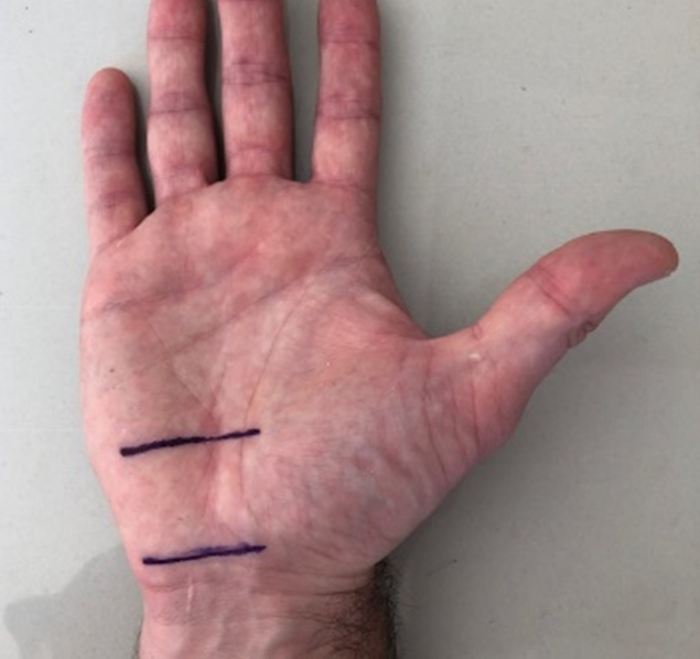
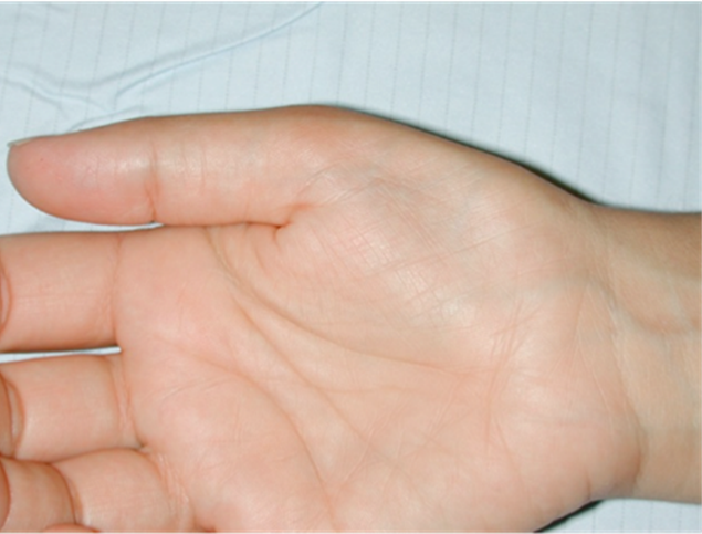
Look for wasting of the thenar eminence. Sensory testing will be normal in early disease and show decreased sensation in the median innervated fingers later in established carpal tunnel syndrome. Remember the proximal palm is innervated by the superficial palmar branch and should not be involved as this branch of the median nerve does not pass through the carpal tunnel. Test thumb abduction power, specifically noting abductor pollicis brevis contraction.
Special tests
- Tinel - tapping over the course of the median nerve will produce tingling in the median nerve distribution when the tapping occurs over or just proximal to the carpal tunnel.
- Phalen - Prolonged deep flexion of the wrist will reproduce symptoms after 30 - 60 seconds.
- Durkan (most sensitive test) - digital compression over the carpal tunnel reproduces symptoms after 30 - 60 seconds.
Special investigations
The diagnosis is clinical and can often be suspected on history alone, and therefore special investigations are not necessary. In atypical cases, nerve conduction studies (NCS) or electromyography (EMG) can be requested.
Imaging
Imaging studies have a limited role in the diagnosis unless carpal tunnel compression secondary to other pathology is suspected.
Management
Patients with associated medical problems should first be managed medically, especially those with hypothyroidism. Pregnant patients should be managed conservatively where possible as the disease often resolves postpartum.
Non-surgical
Mild disease is when there are only intermittent symptoms such as at night or only with certain activities.
Moderate disease is present in those with persistent objective sensory changes but no motor weakness.
Severe disease is considered when motor weakness is evident, and sensation is severely decreased or absent. It is important to note that severe disease is often pain-free due to almost complete sensory loss. A factor that unfortunately further delays presentation as patients think that reduction in pain is evidence of improvement.
Splinting at night works especially well in mild disease with night-time symptoms only. Full time splinting can be used in pregnancy.
Steroid injection into the carpal tunnel can relieve symptoms, but usually only provides temporary relief and risks intraneural injection with nerve damage and chronic pain. A good response to an injection is a good prognostic indicator that surgery will be successful.
The hand therapist has a role in conservative management such as when ergonomic factors are thought to be contributory, or custom splints are required.
Surgical
Patients who have failed conservative measures, have constant symptoms, wasting or weakness should be referred for carpal tunnel release. Patients with symptoms lasting longer than six months have a significantly poorer response to steroid injection and should also be referred. Surgery entails simple division of the transverse carpal ligament. An examination of the carpal tunnel at the time of surgery excludes any secondary causes such as significant synovitis and masses such as ganglions and so on.
Complications
Severe established carpal tunnel syndrome can lead to permanent symptoms of numbness, paraesthesia, weakness and even neuropathic pain.
Carpal tunnel surgery is very successful, but complications can occur, such as persistent pain at the incision area known as pillar pain. The syndrome can also reoccur.
Pitfalls
- Missing the underlying condition that may be present as the cause of carpal tunnel syndrome.
- Misdiagnosing C6 radiculopathy or peripheral neuropathy as carpal tunnel syndrome.
- Misdiagnosing cubital tunnel syndrome (ulnar nerve compression at the elbow) as carpal tunnel syndrome.
References
Hammert WC, Calfee RP, Bozentka DJ, Boyer MI, 2010. ASSH Manual of Hand Surgery; Lippincott, Williams and Wilkins (Wolters Kluwer); Philadelphia, USA.
Miller MD, 2008. Review of Orthopaedics, 5th edition; Elsevier; Philadelphia, USA.
Wolfe SW, Hotchkiss RN, Pederson WC, Kozin SH, Cohen MS, 2017. Green’s Operative Hand Surgery, 7th Edition; Elsevier; Philadelphia, USA.
Assessment
A 60-year-old female presents complaining of waking early in the mornings with a swollen numb hand and having to shake it out. What is your next management step?
- Urgent nerve conduction studies.
- Refer for urgent surgery.
- Prescribe NSAIDs and tell her it will get better.
- Refer to the hand therapist for a splint.
The correct answer is (D), refer to the hand therapist for a splint.